THP-1 Xenograft Model Overview
The THP-1 xenograft model is derived from a human monocytic leukemia cell line established from the peripheral blood of a 1-year-old male patient with acute monocytic leukemia (AML M5). This model is widely recognized for its close resemblance to primary monocytic leukemia blasts, both morphologically and phenotypically, and has become a key tool in studying inflammation, innate immune signaling, and drug responses in monocytic AML. THP-1 xenografts are particularly valuable for investigating agents targeting the NF-κB pathway, TLR signaling, inflammasome activity, and myeloid cell differentiation. Its robust response to phorbol esters and capacity for differentiation into macrophage-like cells add further relevance to its use in both oncology and immunology-focused preclinical studies.
Request a Custom Quote for THP-1 Xenograft ModelBiological and Molecular Characteristics
THP-1 cells are suspension cells that exhibit round to oval morphology with modest cytoplasmic granularity. The cells express markers characteristic of monocyte lineage, including CD11b, CD14, CD33, CD68, and HLA-DR, while lacking lymphoid and granulocytic markers. THP-1 cells carry a t(9;11)(p22;q23) chromosomal translocation that results in the MLL-AF9 fusion gene, a known driver of monocytic leukemia and a poor prognostic factor in AML. They are p53 wild-type and show constitutive activation of NF-κB and STAT pathways. The line is also frequently used to model innate immune responses due to high expression of TLR4 and NLRP3 inflammasome components, and it can be induced to differentiate into adherent, macrophage-like cells upon PMA stimulation.
| Characteristic | THP-1 Cell Line Profile |
|---|---|
| Disease Origin | Acute monocytic leukemia (AML M5) |
| Key Translocation | t(9;11)(p22;q23), MLL-AF9 fusion |
| Immunophenotype | CD11b⁺, CD14⁺, CD33⁺, HLA-DR⁺, CD68⁺ |
| Signaling Pathways | NF-κB, STAT, TLR, NLRP3 |
| Differentiation Capacity | High (macrophage induction via PMA) |
| p53 Status | Wild-type |
In Vivo Model Development and Tumorigenicity
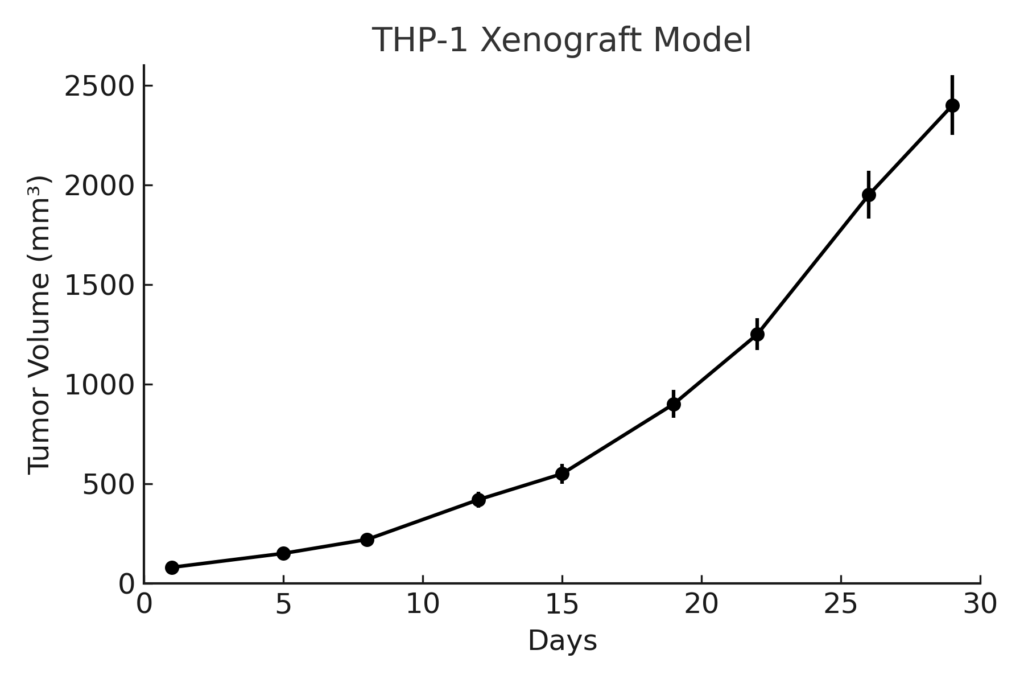
THP-1 xenografts are typically established via subcutaneous or intravenous injection into immunodeficient mice such as NOD/SCID or NSG strains. Subcutaneous models develop localized tumors that reach measurable volumes between 700–900 mm³ within 4–5 weeks and allow for reproducible volume-based pharmacodynamic and efficacy studies. Systemic models, though less common, can be developed via intravenous injection and result in infiltration of bone marrow, spleen, and liver. The tumor take rate is high, particularly when using Matrigel co-injection in subcutaneous studies. The THP-1 model is frequently used to evaluate small molecules, cytokine inhibitors, immune checkpoint modulators, and myeloid differentiation therapies.
Request a Custom Quote for THP-1 Xenograft ModelHistopathology and Immunohistochemical Profile
Histological sections of THP-1 xenografts reveal dense sheets of medium to large monoblasts with vesicular nuclei, occasional nucleoli, and abundant pale cytoplasm. Hematoxylin and eosin staining highlights tumor uniformity with limited necrosis in early stages. Immunohistochemically, xenografts show robust expression of CD68, CD14, and CD33, confirming monocytic lineage. Ki-67 staining indicates high proliferative capacity, and phosphorylated NF-κB and STAT3 are frequently detected in nuclear compartments. Macrophage markers may also be observed when differentiation-inducing treatments are applied in vivo. These features support the model’s use in both leukemic proliferation and innate immune activation studies.
Preclinical Applications and Drug Response
The THP-1 xenograft model is widely used to evaluate therapies targeting monocytic AML, including FLT3 inhibitors, bromodomain (BET) inhibitors, and HDAC inhibitors. It has also been employed to study immune-modulating agents such as IL-1β inhibitors, caspase-1 blockers, and TLR4 antagonists. The model’s sensitivity to NF-κB and inflammasome modulation makes it especially valuable for drug discovery efforts focused on inflammation-associated leukemias. THP-1 also supports testing of pro-differentiation regimens, chemotherapeutics, and novel antibody therapies directed at CD33 or CD123. As such, the model enables comprehensive in vivo evaluation of both anti-leukemic efficacy and host immune interactions.
Request This Model
To incorporate the THP-1 xenograft model into your leukemia or immuno-oncology research program, connect with our scientific team for detailed model specifications and tailored support in designing subcutaneous or systemic in vivo efficacy studies that address the unique pathophysiology of monocytic AML.
Request a Custom Quote for THP-1 Xenograft Model