PC9 Xenograft Model Overview
The PC9 xenograft model originates from a human non-small cell lung carcinoma (NSCLC) cell line derived from a lung adenocarcinoma patient. PC9 cells are characterized by a sensitizing mutation in the epidermal growth factor receptor (EGFR) gene, making this model one of the most widely studied platforms for EGFR-targeted therapy evaluation. The PC9 cell line harbors an exon 19 deletion (delE746-A750) in the EGFR gene, a mutation associated with favorable clinical response to first-generation tyrosine kinase inhibitors (TKIs). The PC9 xenograft model is extensively used to evaluate mechanisms of drug sensitivity, resistance development, and combination strategies in preclinical NSCLC studies.
Request a Custom Quote for PC9 Xenograft ModelBiological and Molecular Characteristics
PC9 cells display classical epithelial morphology and a well-differentiated adenocarcinoma phenotype. The hallmark EGFR exon 19 deletion results in constitutive receptor activation and downstream signaling through the PI3K/AKT and RAS/RAF/MEK/ERK pathways. The cell line is PTEN-positive and wild-type for KRAS, ALK, and BRAF, making it an ideal model for isolating EGFR-dependent oncogenic signaling. PC9 cells exhibit high expression of epithelial markers such as E-cadherin and cytokeratin 7 (CK7), and lack mesenchymal marker expression. These properties enable consistent and reproducible in vivo tumor formation, as well as fidelity in replicating drug responses observed in EGFR-mutant NSCLC patients.
| Characteristic | PC9 Cell Line Profile |
|---|---|
| Cancer Type | Non-small cell lung carcinoma (adenocarcinoma) |
| EGFR Mutation | Exon 19 deletion (delE746-A750) |
| PTEN Status | Wild-type |
| KRAS/BRAF/ALK Status | Wild-type |
| Dominant Pathways | EGFR, PI3K/AKT, MEK/ERK |
| Marker Expression | E-cadherin⁺, CK7⁺, Cytokeratin⁺ |
In Vivo Model Development and Tumorigenicity
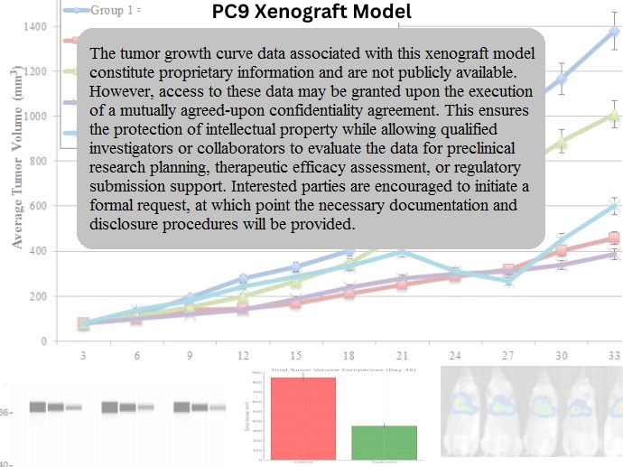
Subcutaneous xenografts are established by injecting PC9 cells into immunodeficient mice, such as nude or NSG strains. Tumors typically become palpable within 7–10 days, with volumes reaching 600–900 mm³ by 4–5 weeks post-inoculation. PC9 xenografts display rapid and homogeneous tumor growth, making them suitable for longitudinal treatment studies and pharmacokinetic-pharmacodynamic evaluations. The model has a high engraftment rate and is compatible with tumor regression protocols under TKI administration. While most studies utilize the subcutaneous format, orthotopic or metastatic models may be developed for studying tumor progression or drug penetration in the pulmonary microenvironment.
Request a Custom Quote for PC9 Xenograft ModelHistopathology and Immunohistochemical Profile
Histologically, PC9 xenograft tumors present as moderately to well-differentiated adenocarcinomas, consisting of uniform epithelial cells forming glandular and solid structures. The tumor cells exhibit round nuclei, finely granular chromatin, and occasional mitotic figures. Immunohistochemistry reveals strong membrane-localized EGFR expression and robust Ki-67 staining, indicative of active proliferation. Additional markers such as phospho-EGFR, phospho-AKT, and phospho-ERK are upregulated, reflecting the active signaling state of the tumor. Staining for epithelial markers, including CK7 and E-cadherin, supports the maintenance of the adenocarcinoma phenotype in vivo.
Preclinical Applications and Drug Response
The PC9 xenograft model is the gold standard for evaluating sensitivity to first- and second-generation EGFR inhibitors, including gefitinib, erlotinib, and afatinib. It is also commonly used to assess the impact of acquired resistance mutations, such as T790M, by introducing derivative sublines or inducing resistance in vivo. The model supports combinatorial drug testing, particularly strategies involving dual inhibition of EGFR and compensatory signaling nodes (e.g., PI3K, MEK). It is frequently applied in translational studies aimed at biomarker validation, dose optimization, and elucidation of EGFR signaling dynamics under therapeutic pressure.
Request This Model
To incorporate the PC9 xenograft model into your NSCLC drug development program or biomarker discovery studies, contact our team for detailed model specifications, tumor kinetics data, and custom study design services tailored to EGFR-driven lung cancer research.
Request a Custom Quote for PC9 Xenograft Model