NCI-H1568 Xenograft Model Overview
The NCI-H1568 xenograft model is derived from a human lung adenocarcinoma and represents a valuable tool for investigating the biology and treatment of non-small cell lung cancer (NSCLC) driven by KRAS and TP53 mutations. Originating from a male patient’s pleural effusion, the NCI-H1568 cell line has been extensively used in translational oncology to evaluate therapeutic agents targeting signaling pathways implicated in treatment-resistant lung cancer. When implanted subcutaneously into immunodeficient mice, this model forms tumors with high reproducibility and predictable growth kinetics, facilitating controlled preclinical evaluation of cytotoxic and molecularly targeted agents. Its defined oncogenic profile and robust tumorigenicity position it as a preferred system for drug efficacy, resistance mechanism, and combination therapy studies in KRAS-driven NSCLC.
Request a Custom Quote for NCI‑H1568 Xenograft ModelBiological and Molecular Characteristics
NCI-H1568 cells exhibit phenotypic and genotypic features reflective of aggressive lung adenocarcinoma. The cell line is characterized by activating KRAS mutations and dysfunctional TP53, which jointly drive oncogenesis through constitutive activation of the MAPK and PI3K pathways and impaired apoptosis. These features contribute to resistance against EGFR inhibitors and sensitivity to agents targeting downstream signaling intermediates. Immunophenotypically, the cells express epithelial markers including cytokeratin 7 and E-cadherin, consistent with their origin. PD-L1 expression is low to moderate, enabling studies focused on checkpoint blockade sensitization. This molecular composition provides a well-characterized foundation for therapeutic screening, especially for agents designed to disrupt oncogenic RAS signaling or restore apoptotic responses.
| Characteristic | Description |
|---|---|
| Tissue Origin | Human lung adenocarcinoma (pleural effusion) |
| Key Mutations | KRAS (activating), TP53 (loss-of-function) |
| Cell Morphology | Epithelial, adherent |
| Immunomarkers | CK7+, E-cadherin+, PD-L1 (low-moderate) |
| Oncogenic Pathways | MAPK, PI3K/AKT, apoptotic dysregulation |
In Vivo Model Development and Tumorigenicity
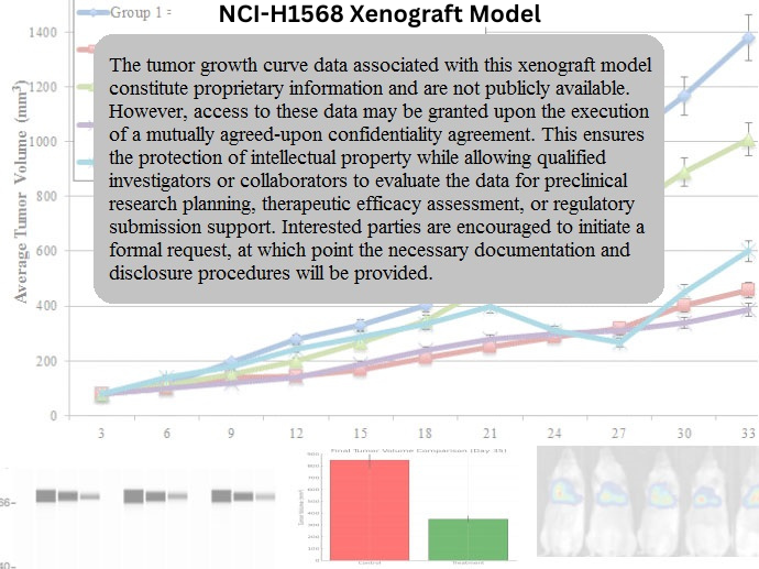
The NCI-H1568 xenograft model is established through subcutaneous injection into immunodeficient mice, including nude or NOD/SCID strains. Tumors typically form within 10–14 days and grow consistently to volumes of 300–500 mm³ within 4–5 weeks, allowing for defined treatment windows and endpoint planning. The model is particularly suited for evaluating inhibitors of RAS-effector pathways and therapeutic combinations designed to overcome intrinsic resistance. Tumor vascularization supports reliable drug delivery, and the reproducible growth kinetics facilitate longitudinal pharmacodynamic studies. The model’s compatibility with imaging modalities, including bioluminescent or PET-based tracking when transduced, further enhances its utility in complex study designs.
Request a Custom Quote for NCI‑H1568 Xenograft ModelHistopathology and Immunohistochemical Profile
Tumors derived from NCI-H1568 xenografts exhibit histopathological hallmarks of moderately differentiated adenocarcinoma. Hematoxylin and eosin staining reveals glandular structures embedded in fibrous stroma, with nuclear pleomorphism and high mitotic activity. Ki-67 staining demonstrates a proliferative index typically exceeding 60%, highlighting the model’s suitability for proliferation-based drug assessments. Immunohistochemical profiling confirms epithelial lineage through cytoplasmic CK7 and membranous E-cadherin expression. Mutant p53 protein accumulation is commonly observed, in line with its nonfunctional status. PD-L1 staining varies across tumor sections but remains relatively low, supporting its relevance in preclinical studies of immunotherapy enhancement strategies.
Preclinical Applications and Drug Response
The NCI-H1568 xenograft model has been widely applied in studies aimed at overcoming KRAS-driven therapeutic resistance. It is resistant to first-generation EGFR inhibitors, making it ideal for evaluating alternative signaling inhibitors such as MEK, ERK, or PI3K-targeted compounds. Combination therapies, particularly those incorporating BCL-2/BCL-XL inhibitors or chemotherapeutics such as docetaxel or pemetrexed, have demonstrated enhanced efficacy in this system. Additionally, the model is useful for assessing delivery technologies, including liposomal and nanoparticle formulations, due to its predictable tumor architecture. The low baseline PD-L1 expression facilitates studies of immunomodulatory agents designed to upregulate immune visibility or enhance T-cell infiltration, particularly in synergy with targeted inhibitors.
Request This Model
To request the NCI-H1568 xenograft model for your preclinical studies, please use the form below. A customized quote and additional model specifications will be provided upon inquiry.
Request a Custom Quote for NCI‑H1568 Xenograft Model