MEG-01 Xenograft Model Overview
The MEG-01 xenograft model originates from a human chronic myeloid leukemia (CML) cell line that was established from the bone marrow of a 5-year-old male patient in the blast crisis phase of CML. MEG-01 cells exhibit megakaryoblastic features and express a variety of platelet-specific markers, providing a unique system to study the biology of megakaryocyte differentiation and thrombopoiesis in the context of leukemic transformation. Importantly, MEG-01 cells harbor the BCR-ABL1 fusion gene resulting from the Philadelphia chromosome translocation t(9;22)(q34;q11), which drives constitutive tyrosine kinase signaling and leukemogenesis. The MEG-01 xenograft model supports in vivo investigation of BCR-ABL–targeted therapies, megakaryocytic dysregulation, and mechanisms underlying blast phase progression in CML.
Request a Custom Quote for MEG‑01 Xenograft ModelBiological and Molecular Characteristics
MEG-01 cells display spontaneous differentiation toward the megakaryocytic lineage and produce platelet-like particles in vitro. Their immunophenotype includes expression of CD41 (GPIIb), CD42b (GPIbα), CD61 (GPIIIa), and CD36, with low to moderate expression of CD34, indicating a transition from progenitor to mature megakaryocyte phenotype. The presence of the BCR-ABL1 fusion gene results in constitutive activation of the ABL tyrosine kinase domain, leading to hyperactivation of downstream pathways including PI3K/AKT, MAPK/ERK, and STAT5. MEG-01 cells are sensitive to tyrosine kinase inhibitors (TKIs) such as imatinib, nilotinib, and dasatinib, though they serve as an effective model for exploring resistance mechanisms and blast crisis biology in CML.
| Characteristic | MEG-01 Cell Line Profile |
|---|---|
| Disease Origin | Chronic myeloid leukemia (blast crisis) |
| Lineage | Megakaryoblastic |
| Key Genetic Alteration | BCR-ABL1 fusion (t(9;22)(q34;q11)) |
| Immunophenotype | CD41⁺, CD42b⁺, CD61⁺, CD36⁺, CD34⁻/low |
| Differentiation Status | Pro-megakaryocytic to mature |
| Therapeutic Sensitivity | TKIs (imatinib, dasatinib), JAK/STAT pathway agents |
In Vivo Model Development and Tumorigenicity
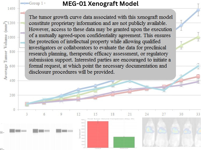
The MEG-01 xenograft model is typically established via subcutaneous injection of cells into immunocompromised mice, including NOD/SCID or NSG strains. Tumor take rates are high, and nodules reach measurable size within 3 to 4 weeks, with volumes reaching 700–900 mm³ by study endpoint. The subcutaneous model offers a robust and controllable environment for evaluating tumor progression, drug efficacy, and pharmacodynamic endpoints. Although systemic models via intravenous injection have been attempted, they yield lower engraftment efficiency compared to other leukemic lines. For platelet biology and megakaryocyte maturation studies, MEG-01 xenografts provide a tractable system for evaluating differentiation-inducing agents and megakaryopoiesis-targeted therapies.
Request a Custom Quote for MEG‑01 Xenograft ModelHistopathology and Immunohistochemical Profile
Histological evaluation of MEG-01 xenografts reveals sheets of large, irregularly shaped cells with lobulated or multilobed nuclei, abundant eosinophilic cytoplasm, and evidence of cytoplasmic granulation, reflecting megakaryocytic differentiation. Hematoxylin and eosin staining highlights minimal necrosis and moderate mitotic activity. Immunohistochemical analysis confirms expression of CD41, CD42b, and CD61 on cell membranes and in the cytoplasm. Proliferation is evidenced by moderate-to-high Ki-67 positivity, while phosphorylated STAT5 and ERK serve as markers of active BCR-ABL signaling. In treated tumors, a decrease in phosphorylated ABL and downstream effectors can serve as pharmacodynamic readouts of TKI activity.
Preclinical Applications and Drug Response
The MEG-01 xenograft model is widely utilized for evaluating BCR-ABL–targeted tyrosine kinase inhibitors, as well as combination therapies aimed at overcoming resistance in CML blast crisis. It is also employed to assess agents targeting megakaryocyte differentiation, apoptosis regulation, and thrombopoiesis-related signaling. Studies investigating the modulation of STAT5, PI3K, and JAK pathways have demonstrated therapeutic relevance in MEG-01 tumors. Additionally, this model serves as a surrogate for preclinical assessment of platelet-derived microparticles and their contribution to thrombosis, inflammation, and tumor microenvironment interactions. MEG-01 xenografts thus provide a dual platform for leukemia and megakaryopoiesis research.
Request This Model
To incorporate the MEG-01 xenograft model into your leukemia or megakaryocytic lineage studies, contact our team to obtain detailed model protocols, support with study design, and access to validated tumor banking and analysis services tailored to CML blast phase research.
Request a Custom Quote for MEG‑01 Xenograft Model