MDST8 Xenograft Model Overview
The MDST8 xenograft model is derived from a human colorectal carcinoma cell line established from the primary tumor of a 72-year-old male patient. Known for its high metastatic potential and robust tumorigenicity, MDST8 is particularly useful for preclinical studies focused on metastatic colorectal cancer. This model is ideal for evaluating drug efficacy, particularly in the context of metastatic disease, and is commonly used to study the molecular mechanisms underlying metastasis, drug resistance, and epithelial-mesenchymal transition (EMT) in colorectal carcinoma. MDST8 xenografts provide a stable and reproducible platform for evaluating combination therapies, immunotherapies, and novel agents designed to target the metastatic progression of colorectal cancer.
Request a Custom Quote for MDST8 Xenograft ModelBiological and Molecular Characteristics
MDST8 cells exhibit classic epithelial morphology, with tight cell–cell junctions and a high degree of cell adhesion. The cell line is microsatellite stable (MSS) and carries wild-type KRAS, NRAS, and BRAF, ensuring its responsiveness to EGFR-targeted therapies such as cetuximab and panitumumab. TP53 is mutated in this model, leading to compromised apoptotic regulation and making it a suitable model for evaluating DNA-damaging agents and therapies targeting the DNA repair mechanisms. MDST8 cells express moderate levels of carcinoembryonic antigen (CEA), cytokeratin 20 (CK20), and other markers indicative of colorectal origin. Wnt/β-catenin signaling is also active in MDST8, with β-catenin localized both in the cytoplasm and at the membrane, indicating active signaling through this pathway. These molecular features render MDST8 an excellent system for studying the efficacy of EGFR inhibitors, chemotherapeutic agents, and combination therapies.
| Characteristic | MDST8 Cell Line Profile |
|---|---|
| Tissue of Origin | Colorectal adenocarcinoma (primary) |
| KRAS/NRAS/BRAF Status | Wild-type |
| TP53 Status | Mutated |
| MSI Status | Microsatellite stable (MSS) |
| Differentiation Markers | CK20, CEA, E-cadherin |
| Wnt Signaling | Active, β-catenin cytoplasmic/membranous |
In Vivo Model Development and Tumorigenicity
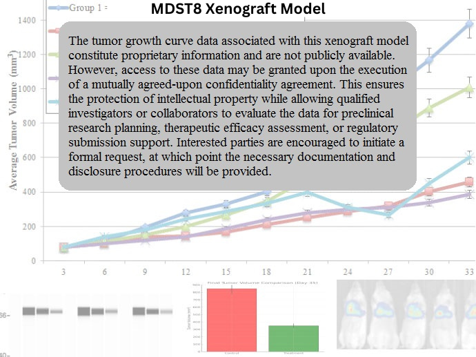
MDST8 xenografts are typically established by subcutaneous or orthotopic injection of cultured cells into immunodeficient mice, such as athymic nude or NOD/SCID strains. Tumor formation is generally rapid, with growth detectable within 7 to 10 days post-inoculation. Tumors typically reach volumes of 700–900 mm³ within 4 to 5 weeks, allowing for robust in vivo studies of drug efficacy and metastasis. The model’s high metastatic potential is particularly evident in the formation of secondary lesions, particularly in the liver and lungs, making it an excellent model for evaluating therapies targeting metastatic progression and invasion. The consistency in tumor growth and metastatic spread provides a reliable platform for long-term studies of drug resistance, tumor recurrence, and treatment regimens involving multiple therapeutic modalities.
Request a Custom Quote for MDST8 Xenograft ModelHistopathology and Immunohistochemical Profile
Histopathologically, MDST8-derived xenografts exhibit moderately differentiated adenocarcinomas with glandular structures, focal areas of necrosis, and moderate mitotic activity. Hematoxylin and eosin (H&E) staining highlights well-polarized epithelial cells with basally oriented nuclei and clear cytoplasm. Immunohistochemical staining confirms strong expression of CEA, CK20, and E-cadherin, validating the colorectal origin of the tumor and its epithelial nature. β-catenin staining shows both membranous and cytoplasmic expression, indicative of active Wnt signaling. Mutant p53 protein is detected in the tumor nuclei, reflecting the loss of apoptotic function in the cells. The model’s ability to develop metastases and invade surrounding tissues makes it particularly valuable for studying the effects of drugs on tumor spread and the underlying molecular mechanisms of metastasis.
Preclinical Applications and Drug Response
The MDST8 xenograft model is highly relevant for evaluating therapies targeting metastatic colorectal cancer, particularly in MSS tumors. Its wild-type KRAS, NRAS, and BRAF background ensures its responsiveness to EGFR inhibitors, such as cetuximab and panitumumab, and makes it a reliable system for studying the effects of these agents on primary and metastatic tumors. The TP53 mutation in this model provides an opportunity to evaluate DNA-damaging agents, as well as therapies that exploit synthetic lethality in tumor cells. MDST8 xenografts are also widely used in studies of metastasis, including evaluating therapies that inhibit tumor migration, invasion, and angiogenesis. The model’s high metastatic potential makes it ideal for combination studies involving chemotherapy, targeted agents, and immunotherapies aimed at overcoming metastasis-driven resistance.
Request This Model
To incorporate the MDST8 xenograft model into your preclinical colorectal cancer research or drug development program, contact our scientific team to discuss model specifications, customized study designs, and access to this valuable metastatic cancer model.
Request a Custom Quote for MDST8 Xenograft Model