KATO-III Xenograft Model Overview
The KATO-III xenograft model is derived from an undifferentiated human gastric carcinoma originally established from a metastatic lymph node of a 55-year-old male patient. It represents a signet-ring cell type gastric cancer, classified under the diffuse subtype according to Lauren’s classification. KATO-III xenografts recapitulate key features of aggressive, poorly cohesive gastric adenocarcinomas with metastatic potential and resistance to standard chemotherapeutics. As a HER2-negative and CDH1-deficient model, KATO-III is well suited for evaluating therapeutic interventions in genomically stable (GS), microsatellite-stable (MSS) gastric tumors that lack well-defined molecular targets. The model’s reproducible tumor growth, high cellular plasticity, and clinical relevance make it a robust system for preclinical oncology research.
Request a Custom Quote for KATO-III Xenograft ModelBiological and Molecular Characteristics
KATO-III cells display classic signet-ring morphology with intracytoplasmic mucin vacuoles and crescent-shaped nuclei. The cell line is negative for HER2 amplification and deficient in CDH1, the gene encoding E-cadherin, leading to impaired cell adhesion and increased invasiveness. KATO-III is wild-type for KRAS and BRAF, and lacks mismatch repair deficiency, confirming a microsatellite-stable (MSS) phenotype. TP53 is mutated, and the PI3K/AKT and JAK/STAT3 pathways are activated, supporting tumor cell survival, proliferation, and chemoresistance. The loss of tight junction proteins and expression of mesenchymal markers such as vimentin and ZEB1 further implicate epithelial–mesenchymal transition (EMT) in the tumor biology of this model.
| Characteristic | KATO-III Cell Line Profile |
|---|---|
| Tissue of Origin | Gastric signet-ring cell carcinoma (metastatic) |
| HER2 Status | Negative |
| CDH1 Status | Deficient |
| KRAS/BRAF Status | Wild-type |
| TP53 Status | Mutated |
| MSI Status | Microsatellite stable (MSS) |
| EMT Markers | ↑Vimentin, ↑ZEB1, ↓E-cadherin |
In Vivo Model Development and Tumorigenicity
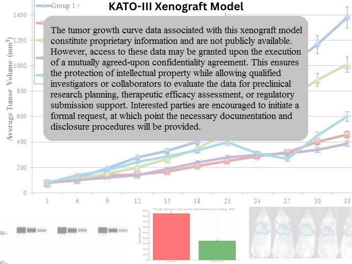
KATO-III xenografts are typically established through subcutaneous injection of tumor cells into immunodeficient mice, such as athymic nude or NOD/SCID strains. Tumors exhibit a moderate to aggressive growth profile, with initial engraftment occurring within 10–14 days and progression to 700–900 mm³ by 4–5 weeks. Due to their diffuse cellular characteristics and loss of adhesion, KATO-III xenografts often display a non-cohesive growth pattern, mimicking the infiltrative nature of signet-ring cell carcinomas. This model is particularly suited for studying tumor invasiveness, peritoneal dissemination, and the therapeutic targeting of EMT-associated pathways. It provides a relevant system for evaluating both standard-of-care chemotherapies and novel anti-metastatic agents.
Request a Custom Quote for KATO-III Xenograft ModelHistopathology and Immunohistochemical Profile
Histological analysis of KATO-III xenograft tumors reveals a diffuse infiltrative pattern composed of signet-ring cells dispersed through the stromal matrix. Hematoxylin and eosin (H&E) staining highlights intracytoplasmic mucin and eccentric nuclei, with minimal glandular architecture. Immunohistochemically, tumors demonstrate strong nuclear p53 expression due to TP53 mutation and absent E-cadherin staining, consistent with CDH1 deficiency. Vimentin and ZEB1 expression is elevated, supporting the mesenchymal phenotype. HER2 staining is consistently negative, further defining the tumor as a non-amplified gastric adenocarcinoma. The pathological features of KATO-III xenografts align closely with advanced diffuse gastric cancer and provide high translational value.
Preclinical Applications and Drug Response
The KATO-III xenograft model is utilized in a range of preclinical applications focusing on aggressive gastric cancers with poor therapeutic response. It is particularly useful in testing chemotherapeutic combinations involving 5-fluorouracil, platinum compounds, and irinotecan. Owing to its EMT features, the model supports evaluation of inhibitors targeting TGF-β, STAT3, and PI3K/AKT pathways, as well as agents aimed at reversing mesenchymal transition. Additionally, KATO-III is frequently used in studies of anti-metastatic compounds, extracellular matrix modulators, and immunotherapeutic strategies in HER2-negative gastric cancer. The model’s molecular stability and clinical mimicry make it a valuable tool for dissecting drug resistance and metastatic behavior in gastric carcinoma.
Request This Model
To integrate the KATO-III xenograft model into your gastric cancer research or therapeutic development pipeline, reach out to our scientific team for full model specifications and assistance with tailored study design for signet-ring and diffuse-type gastric adenocarcinomas.
Request a Custom Quote for KATO-III Xenograft Model