HuTu80 Xenograft Model Overview
The HuTu80 xenograft model is derived from a human colorectal adenocarcinoma cell line established from a primary tumor in a 63-year-old male patient. Known for its well-differentiated epithelial morphology and robust tumorigenic potential, HuTu80 is frequently used in preclinical studies focused on colorectal cancer, particularly for evaluating novel therapeutic agents, drug resistance mechanisms, and the effects of EGFR-targeted therapies. This model’s stable growth and reproducibility in vivo make it an ideal candidate for long-term studies, including those aimed at investigating combination therapies, targeted treatments, and the underlying molecular biology of colorectal cancer progression.
Request a Custom Quote for HuTu80 Xenograft ModelBiological and Molecular Characteristics
HuTu80 cells exhibit epithelial morphology with tight cell–cell junctions and high levels of E-cadherin expression, supporting its use in studies of cell adhesion, differentiation, and epithelial integrity. The model is microsatellite stable (MSS) and retains wild-type KRAS, BRAF, and NRAS, making it responsive to EGFR inhibitors like cetuximab and panitumumab. TP53 is mutated in this model, which results in a compromised apoptotic response and offers an opportunity to study therapies targeting DNA repair mechanisms or apoptosis. HuTu80 cells express moderate levels of carcinoembryonic antigen (CEA) and cytokeratin 20 (CK20), confirming their colorectal origin. Additionally, active Wnt/β-catenin signaling is present, with β-catenin localized both at the membrane and in the cytoplasm. These characteristics make HuTu80 an excellent model for evaluating therapies targeting EGFR, Wnt signaling, and DNA repair pathways in colorectal cancer.
| Characteristic | HuTu80 Cell Line Profile |
|---|---|
| Tissue of Origin | Colorectal adenocarcinoma (primary) |
| KRAS/BRAF/NRAS Status | Wild-type |
| TP53 Status | Mutated |
| MSI Status | Microsatellite stable (MSS) |
| Differentiation Markers | CK20, CEA, E-cadherin |
| Wnt Signaling | Active, β-catenin cytoplasmic/membranous |
In Vivo Model Development and Tumorigenicity
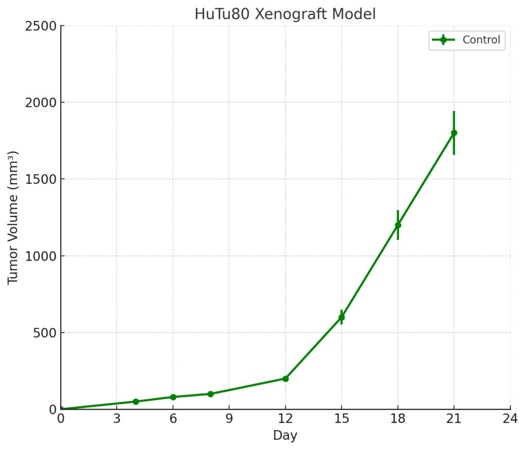
HuTu80 xenografts are typically established by subcutaneous injection of cultured cells into immunodeficient mouse strains such as athymic nude or NOD/SCID mice. Tumor formation occurs within 7 to 10 days post-implantation, with tumors reaching volumes of 700–900 mm³ within 3 to 4 weeks. The model demonstrates consistent tumor growth and a stable histological phenotype, making it suitable for multi-cycle treatment studies and long-term drug efficacy evaluations. Due to its wild-type KRAS and BRAF status, HuTu80 xenografts are particularly useful for testing EGFR inhibitors and combination therapies targeting the EGFR pathway. The model’s TP53 mutation allows for the study of therapies that exploit apoptotic defects or target DNA repair mechanisms in colorectal cancer.
Request a Custom Quote for HuTu80 Xenograft ModelHistopathology and Immunohistochemical Profile
Histologically, HuTu80 xenografts exhibit well-differentiated adenocarcinomas with glandular structures, minimal mucin production, and areas of central necrosis. Hematoxylin and eosin (H&E) staining highlights well-polarized epithelial cells with basally located nuclei and clear cytoplasm. Immunohistochemical analysis confirms strong expression of epithelial markers such as CK20, CEA, and E-cadherin, verifying the colorectal origin of the tumor. β-catenin staining shows both cytoplasmic and membranous localization, indicating active Wnt signaling. Mutant p53 protein is detectable in the tumor nuclei, underscoring the loss of functional p53-mediated apoptotic control. These histological features support the use of HuTu80 xenografts in colorectal cancer research and therapeutic testing.
Preclinical Applications and Drug Response
The HuTu80 xenograft model is highly suitable for evaluating therapies in KRAS/BRAF wild-type, microsatellite-stable colorectal cancer. The model’s responsiveness to EGFR-targeted therapies such as cetuximab and panitumumab makes it ideal for studying the effects of these agents in both primary tumors and metastatic disease. The TP53 mutation also makes the model useful for evaluating DNA-damaging agents and therapies targeting DNA repair and apoptosis. In addition, HuTu80 xenografts are valuable for testing combination therapies aimed at overcoming resistance to EGFR inhibition, including combinations with Wnt pathway inhibitors, chemotherapy agents, and immune-modulating treatments. The model’s consistent growth and molecular stability allow for in-depth studies of drug efficacy, resistance mechanisms, and biomarker development in colorectal cancer.
Request This Model
To explore the potential of the HuTu80 xenograft model for your colorectal cancer research or drug development program, contact our scientific team for detailed model specifications, customized study designs, and access to this reliable and reproducible model system.
Request a Custom Quote for HuTu80 Xenograft Model