HT1376 Xenograft Model Overview
The HT1376 xenograft model is derived from a grade III transitional cell carcinoma of the human urinary bladder and represents a robust, moderately aggressive model for investigating advanced urothelial carcinoma. Originally isolated from a 58-year-old female patient, the HT1376 cell line exhibits reproducible tumorigenicity, intermediate proliferation kinetics, and well-characterized molecular alterations associated with cell cycle deregulation and oncogenic stress. In vivo, HT1376 tumors develop with consistent latency and present as cohesive, non-metastatic masses, making this model ideal for evaluating therapeutic strategies targeting DNA replication stress, epigenetic dysregulation, and metabolic reprogramming in high-grade bladder cancer.
Unlike other more mesenchymal bladder cancer models, HT1376 retains a mixed epithelial profile with partial urothelial differentiation, allowing for the study of tumors in a biologically transitional state. It is frequently employed in the preclinical development of agents targeting p53-deficient cancers, cell cycle checkpoint inhibitors, and agents that modulate chromatin remodeling, including histone deacetylase and methyltransferase inhibitors. Its reproducible growth patterns and moderate aggressiveness make HT1376 well suited for mechanistic, pharmacologic, and drug resistance studies.
Request a Custom Quote for HT1376 Xenograft ModelBiological and Molecular Characteristics
The HT1376 cell line is characterized by a mutated TP53 gene, leading to non-functional accumulation of p53 protein and downstream impairment of apoptosis and DNA damage response pathways. The cell line also exhibits partial loss of RB1 function and disrupted regulation of cyclin D1 and CDK4, resulting in G1/S checkpoint bypass and unscheduled cell cycle progression. Importantly, HT1376 lacks FGFR3 alterations, distinguishing it from luminal-type bladder cancer models and aligning it more closely with the basal-squamous molecular subtype.
HT1376 cells express E-cadherin, cytokeratin 7, and cytokeratin 18, confirming epithelial lineage, but lack uroplakin II and CK20, indicating incomplete terminal differentiation. Additionally, the cells express MMP-9 and VEGF-A, suggestive of an invasive but non-metastatic phenotype in vivo. The basal activation of PI3K/AKT and ERK1/2 signaling pathways contributes to enhanced survival and proliferation under nutrient-limited conditions.
The table below summarizes key biological and molecular features of the HT1376 cell line:
| Characteristic | HT1376 Profile |
|---|---|
| Origin | Human urinary bladder, grade III carcinoma |
| TP53 Status | Mutated (accumulation of non-functional p53) |
| RB1 Pathway | Partially disrupted |
| FGFR3 Status | Wild-type |
| EGFR Expression | Moderate |
| Differentiation Markers | CK7+, CK18+, E-cadherin+ |
| Terminal Differentiation Markers | Uroplakin II-, CK20- |
| EMT Profile | Absent; epithelial predominant |
| Angiogenic Markers | VEGF-A+, MMP-9+ |
| Growth Factor Pathways | Activated PI3K/AKT, ERK1/2 |
This unique molecular profile provides a reliable experimental platform for testing compounds that target proliferative stress, DNA repair deficiency, and therapeutic vulnerability in epithelial bladder tumors with impaired apoptotic control.
In Vivo Model Development and Tumorigenicity
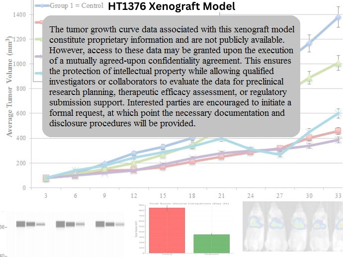
HT1376 xenografts reliably establish tumors in immunodeficient murine hosts, with tumor take rates consistently exceeding 85% in athymic nude or NOD-SCID mice. Subcutaneous implantation of 5 × 10^6 to 1 × 10^7 cells suspended in a 1:1 Matrigel:PBS mixture results in palpable tumor formation within 12–16 days. Tumor growth follows a sigmoidal curve, with endpoint volumes of 1,200–1,400 mm³ typically reached between 28 and 35 days post-implantation.
Tumors derived from HT1376 are non-metastatic under standard subcutaneous conditions and exhibit low local invasion. However, orthotopic delivery methods, including bladder wall injection, have been used to replicate the anatomical context of urothelial carcinoma and assess stromal interaction. The model is compatible with both bioluminescent and fluorescent tagging to support non-invasive imaging and longitudinal tumor tracking.
Its moderate growth rate and consistent tumor architecture make HT1376 suitable for studies that require repeat measurements, time-course sampling, or extended treatment schedules. The model also supports pharmacokinetic and pharmacodynamic profiling, as its vascular density and epithelial compactness enable adequate drug perfusion and tumor tissue penetration.
Request a Custom Quote for HT1376 Xenograft ModelHistopathology and Immunohistochemical Profile
Histologically, HT1376 xenografts display a moderately differentiated transitional cell carcinoma morphology with solid nests of polygonal cells, moderate nuclear pleomorphism, and distinct nucleoli. Tumors retain areas of epithelial polarity, with preserved cell junctions and limited necrosis. Mitoses are frequent but not excessive, and the tumors exhibit low stromal content, resulting in dense cellularity and homogeneous tissue distribution.
Immunohistochemically, HT1376 xenografts demonstrate strong nuclear Ki-67 expression with a labeling index of 50–60%, indicating moderate proliferative capacity. Staining for E-cadherin, CK7, and CK18 confirms epithelial identity, while uroplakin II and CK20 are absent. EGFR is expressed at intermediate levels with membranous localization, while phospho-AKT and phospho-ERK1/2 demonstrate robust cytoplasmic activity.
VEGF-A and MMP-9 are expressed throughout the tumor mass, particularly at invasive margins, supporting angiogenesis and matrix remodeling. CD31-positive vasculature is moderately abundant and evenly distributed. Nuclear accumulation of non-functional p53 confirms the mutation-induced loss of apoptotic regulation, a defining hallmark of this model.
Preclinical Applications and Drug Response
The HT1376 xenograft model is widely utilized for investigating therapies aimed at restoring apoptotic sensitivity in p53-deficient tumors, inhibiting cell cycle progression, or modifying chromatin accessibility through epigenetic drugs. Due to its defective p53 function and partial RB1 disruption, the model is partially resistant to genotoxic chemotherapies such as cisplatin and mitomycin C, making it valuable for identifying mechanisms of resistance and testing agents that overcome checkpoint bypass.
HT1376 has been used in the development and testing of HDAC inhibitors, EZH2 inhibitors, and BRD4 inhibitors, with notable modulation of gene expression, tumor growth suppression, and restoration of epithelial features. Inhibitors of PI3K, AKT, and MEK also show partial activity, especially in combination settings where synthetic lethality can be exploited.
Additionally, the model has been employed in metabolic vulnerability studies, including glycolysis inhibition and autophagy modulation, due to its dependence on PI3K/AKT signaling under stress. HT1376 is particularly suitable for preclinical studies requiring moderate tumor growth, p53-null background, and histologically defined epithelial architecture.
Request This Model
To request the HT1376 xenograft model or to discuss integration into your experimental study design—including drug resistance evaluation, apoptosis restoration, or epigenetic screening—please reach out using the quote request form below. Our team will support all aspects of model acquisition and implementation.
Request a Custom Quote for HT1376 Xenograft Model