HPAF-II Xenograft Model Overview
The HPAF-II xenograft model is derived from a human pancreatic adenocarcinoma cell line, HPAF-II, which was established from a patient with advanced pancreatic cancer. Pancreatic cancer is one of the most lethal cancers, largely due to its late-stage diagnosis, high metastatic potential, and resistance to conventional therapies. The HPAF-II xenograft model is widely used in preclinical research to study the molecular mechanisms of pancreatic cancer progression, evaluate novel therapeutic strategies, and investigate the mechanisms of drug resistance. Given its molecular features and tumorigenic properties, the HPAF-II model is an excellent system for testing chemotherapy, targeted therapies, and immunotherapies.
Request a Custom Quote for HPAF-II Xenograft ModelBiological and Molecular Characteristics
HPAF-II cells are characterized by their epithelial origin and express common markers of pancreatic cancer, including cytokeratins, epithelial membrane antigen (EMA), and the pancreatic cancer-associated antigen CA19-9. The model also exhibits key genetic features, such as mutations in the KRAS gene, which are present in over 90% of pancreatic ductal adenocarcinomas (PDAC) and contribute to tumor initiation, progression, and resistance to treatment. Additionally, HPAF-II cells show loss of the tumor suppressor gene p53, which plays a critical role in regulating the cell cycle and apoptosis. The model also demonstrates dysregulated PI3K/AKT and MAPK signaling pathways, which are frequently activated in pancreatic cancer and contribute to tumor cell survival, proliferation, and metastasis. These characteristics make the HPAF-II xenograft model highly relevant for studying therapies that target these pathways.
| Marker | Expression Level | Function |
|---|---|---|
| Cytokeratin | High | Epithelial cell marker |
| EMA | High | Epithelial membrane antigen |
| CA19-9 | Elevated | Pancreatic cancer biomarker |
| KRAS | Mutated | Oncogene involved in tumor progression |
| PI3K/AKT pathway | Dysregulated | Promotes cell survival and proliferation |
In Vivo Model Development and Tumorigenicity
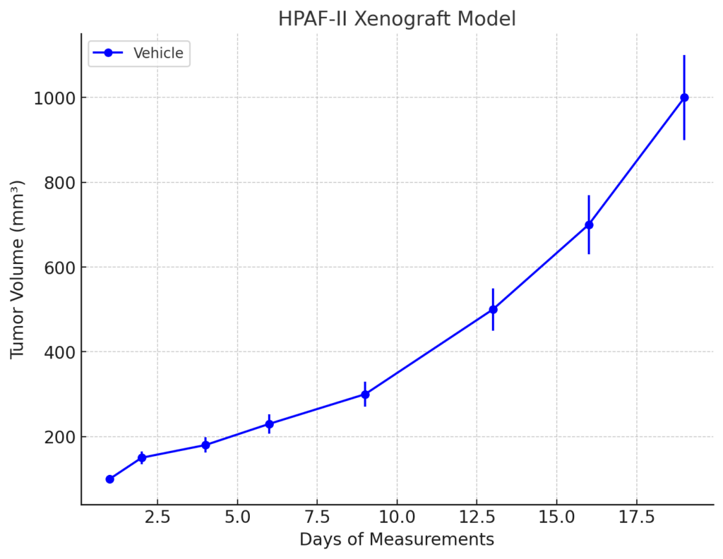
The HPAF-II xenograft model is typically established by subcutaneously implanting HPAF-II cells into immunocompromised mice, such as NOD/SCID or NSG mice. Once implanted, the cells form rapidly growing tumors that replicate the key features of human pancreatic adenocarcinoma, including high cellularity, significant necrosis, and extensive vascularization. The model is widely used to study tumor growth, metastatic spread, and the effects of various therapies. Given its resistance to chemotherapy, including platinum-based agents like gemcitabine, the HPAF-II model is particularly useful for testing combination therapies aimed at overcoming drug resistance or enhancing the effects of standard treatments.
In addition to subcutaneous implantation, orthotopic models of HPAF-II can be established by implanting cells into the pancreas of immunocompromised mice. This method more closely mimics the natural tumor growth and metastasis seen in patients with pancreatic cancer. The orthotopic model allows for the study of tumor progression, invasion, and peritoneal dissemination, providing a more clinically relevant model for testing the effects of new therapies on pancreatic cancer.
Request a Custom Quote for HPAF-II Xenograft ModelHistopathology and Immunohistochemical Profile
Histopathological analysis of HPAF-II xenografts reveals characteristic features of pancreatic adenocarcinoma, including irregular glandular structures and areas of necrosis. The tumors exhibit significant cellular heterogeneity, with both solid and cystic regions present. Immunohistochemical staining of HPAF-II xenografts shows strong expression of epithelial markers, such as cytokeratin and EMA, confirming the epithelial origin of the tumor. Additionally, elevated levels of CA19-9 are observed in the tumor, which is consistent with the biomarker’s role in pancreatic cancer. The tumors also express KRAS, which is mutated in the majority of PDAC cases and is a key driver of tumor progression. Furthermore, HPAF-II xenografts show activation of the PI3K/AKT pathway, as evidenced by high levels of phosphorylated AKT, which contribute to tumor cell survival and resistance to apoptosis. Tumor vascularization, assessed by CD31 staining, indicates the high angiogenic activity within the tumors, supporting their rapid growth.
Preclinical Applications and Drug Response
The HPAF-II xenograft model is widely used to evaluate the efficacy of chemotherapy agents for pancreatic cancer, particularly gemcitabine, which is one of the standard treatments for PDAC. The model is particularly valuable for studying the mechanisms of chemotherapy resistance, as HPAF-II tumors can develop resistance to gemcitabine, providing an excellent system for testing new drugs aimed at overcoming this resistance. Additionally, the model is frequently used to assess targeted therapies, particularly those that inhibit key signaling pathways involved in PDAC progression, such as the KRAS, PI3K/AKT, and MAPK pathways. Given the dysregulated signaling in these pathways, the HPAF-II model is also used to investigate novel therapies targeting these molecular mechanisms.
The HPAF-II xenograft model is also increasingly used to evaluate immunotherapies, including immune checkpoint inhibitors and CAR-T cell therapies, which have shown promise in clinical trials for various cancers. The model’s ability to replicate the immune-suppressive tumor microenvironment of PDAC makes it an ideal platform for testing these new immunotherapeutic approaches. Furthermore, the model can be used to evaluate agents that target the tumor stroma, including fibroblast activation and collagen deposition, which contribute to the aggressive behavior of pancreatic tumors.
Request This Model
To request the HPAF-II xenograft model for your preclinical studies, please use the form below. A customized quote and additional model specifications will be provided upon inquiry.
Request a Custom Quote for HPAF-II Xenograft Model