HEC-1 Xenograft Model Overview
The HEC-1 xenograft model originates from the HEC-1-A and HEC-1-B cell lines, both derived from a human endometrial adenocarcinoma resected from a 71-year-old female patient. The model represents type I endometrial cancer and is widely utilized in preclinical research due to its reproducible tumorigenicity, hormone responsiveness, and molecular fidelity to early-stage, estrogen-influenced malignancies. HEC-1 xenografts are especially valuable for evaluating the effects of hormonal therapy, chemotherapeutic agents, and targeted pathway inhibitors in endometrial carcinoma. Their ability to form histologically accurate tumors in vivo makes them a robust platform for translational research in gynecologic oncology.
Request a Custom Quote for HEC-1 Xenograft ModelBiological and Molecular Characteristics
HEC-1 cells are epithelial in morphology and demonstrate features characteristic of moderately differentiated endometrioid adenocarcinoma. These cells are estrogen receptor (ER)-positive and progesterone receptor (PR)-positive, which supports their use in studies of endocrine regulation. The line is PTEN-deficient, resulting in constitutive activation of the PI3K/AKT/mTOR pathway. It is wild-type for TP53, aligning with the genetic landscape of type I endometrial cancers. Microsatellite instability (MSI) is typically absent, categorizing HEC-1 as microsatellite stable (MSS). Expression of epithelial markers such as cytokeratin 8/18 and E-cadherin further confirms their lineage and differentiation status. These molecular characteristics render the model ideal for studying hormonal influences, receptor-targeted therapies, and signaling pathway dependencies.
| Characteristic | HEC-1 Cell Line Profile |
|---|---|
| Tissue of Origin | Endometrial adenocarcinoma (primary tumor) |
| Hormone Receptor Status | ER-positive, PR-positive |
| TP53 Status | Wild-type |
| PTEN Status | Loss-of-function mutation |
| MSI Status | Microsatellite stable (MSS) |
| Differentiation Markers | CK8/18, E-cadherin |
In Vivo Model Development and Tumorigenicity
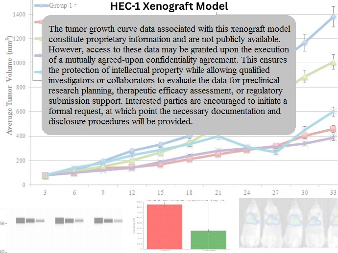
HEC-1 xenografts are developed through subcutaneous implantation of cultured tumor cells into immunocompromised mice, typically nude or NOD/SCID strains. Tumor formation is hormone-sensitive; therefore, estrogen supplementation via subcutaneous pellets is often required for reliable engraftment and sustained growth. Tumors typically form within 10–14 days and progress steadily, reaching volumes of 700–900 mm³ within 4–5 weeks. The model’s consistent tumor take rate and growth kinetics make it suitable for both monotherapy and combination therapy assessments. HEC-1 xenografts are particularly beneficial in evaluating dose-dependent effects of estrogen receptor modulators and inhibitors of PI3K/mTOR signaling in hormone-responsive endometrial cancer.
Request a Custom Quote for HEC-1 Xenograft ModelHistopathology and Immunohistochemical Profile
Histologically, HEC-1 xenograft tumors exhibit glandular structures lined by columnar epithelium with moderate pleomorphism and mitotic activity, reflective of moderately differentiated endometrioid carcinoma. Hematoxylin and eosin (H&E) staining reveals well-formed glands and a pseudostratified nuclear pattern consistent with early-stage endometrial histology. Immunohistochemistry demonstrates strong nuclear staining for ER and PR, confirming the model’s hormone dependence. Expression of CK8/18 and E-cadherin is retained throughout the tumor epithelium, while PTEN loss is evidenced by reduced or absent cytoplasmic staining. This histological and molecular fidelity supports the model’s application in preclinical studies targeting hormone-regulated and growth signaling pathways.
Preclinical Applications and Drug Response
The HEC-1 xenograft model is broadly used for preclinical assessment of hormone-based therapies, including selective estrogen receptor modulators (SERMs), aromatase inhibitors, and anti-progestins. Due to its PTEN deficiency and downstream AKT activation, it is highly responsive to inhibitors of the PI3K/AKT/mTOR axis, making it suitable for testing dual-targeted regimens. The model is also employed in research evaluating cytotoxic agents, differentiation inducers, and endocrine resistance mechanisms in endometrial carcinoma. Its hormone receptor positivity and reproducible tumorigenicity position HEC-1 as an essential system for the development and refinement of therapeutics targeting type I endometrial tumors.
Request This Model
To request access to the HEC-1 xenograft model for your endometrial cancer research or therapeutic development program, contact our scientific team for detailed specifications and customized study planning designed to meet your experimental needs.
Request a Custom Quote for HEC-1 Xenograft Model