HCT-8 Xenograft Model Overview
The HCT-8 xenograft model is derived from a human colorectal carcinoma cell line originally isolated from a metastatic lesion in the peritoneum of a 63-year-old male patient. This model is widely used in preclinical studies focused on advanced colorectal cancer, particularly in metastatic disease models where tumor dissemination and peritoneal spread are key points of interest. The HCT-8 xenograft model is notable for its high tumorigenic potential and consistent growth in vivo, making it a robust platform for evaluating drug efficacy, targeting metastasis, and exploring new treatment strategies for colorectal cancer with aggressive metastatic behavior. HCT-8-derived tumors form peritoneal implants and exhibit features that mimic human metastatic disease, providing a valuable tool for the study of colorectal cancer progression and therapeutic resistance.
Request a Custom Quote for HCT-8 Xenograft ModelBiological and Molecular Characteristics
HCT-8 cells exhibit an epithelial morphology and strong cell–cell adhesion, with key markers such as E-cadherin and cytokeratin 20 (CK20) confirming their colorectal origin. The cell line is microsatellite stable (MSS) and does not harbor KRAS or BRAF mutations, allowing for unimpeded responsiveness to EGFR-targeted therapies. TP53 is mutated in this model, contributing to defective apoptosis and dysregulation of the DNA damage response, which is important for evaluating DNA-damaging agents and treatments that exploit apoptotic defects. Additionally, HCT-8 cells display high levels of carcinoembryonic antigen (CEA) and other markers commonly associated with metastatic colorectal carcinoma. These molecular features make the HCT-8 xenograft an ideal model for studying advanced colorectal cancer therapies, including those targeting EGFR and DNA repair pathways.
| Characteristic | HCT-8 Cell Line Profile |
|---|---|
| Tissue of Origin | Colorectal adenocarcinoma (metastatic) |
| KRAS/BRAF Status | Wild-type |
| TP53 Status | Mutated |
| MSI Status | Microsatellite stable (MSS) |
| Differentiation Markers | CK20, CEA, E-cadherin |
| Tumor Invasion/Metastasis | Peritoneal dissemination, aggressive growth |
In Vivo Model Development and Tumorigenicity
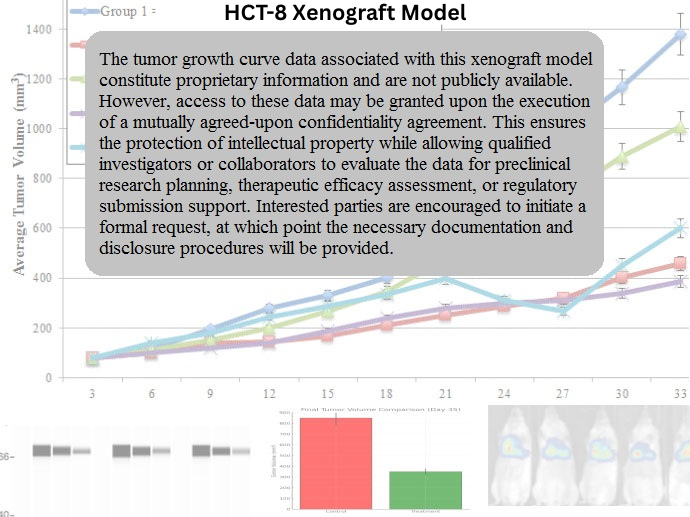
HCT-8 xenografts are established through subcutaneous or intraperitoneal injection of cultured cells into immunodeficient mice, such as athymic nude or NOD/SCID strains. Tumor formation typically occurs within 7–10 days post-implantation, with tumor growth reaching 700–900 mm³ by day 28–35. The model demonstrates aggressive, metastatic spread with the potential for peritoneal dissemination, which is a key feature of advanced colorectal cancer. This model is particularly useful for studying therapies targeting metastatic disease and assessing the effects of drugs on tumor invasion and spread. The consistent growth kinetics and reproducible metastasis make HCT-8 xenografts an ideal system for evaluating multi-agent treatment regimens and exploring novel therapeutic strategies designed to limit metastatic progression in colorectal cancer.
Request a Custom Quote for HCT-8 Xenograft ModelHistopathology and Immunohistochemical Profile
Histopathological analysis of HCT-8 xenografts reveals moderately differentiated adenocarcinomas with invasive growth patterns and peritoneal implants. Hematoxylin and eosin (H&E) staining demonstrates glandular architecture with regions of necrosis, moderate mitotic activity, and peritoneal involvement. The tumors exhibit moderate to high levels of mucin production, consistent with colorectal cancer differentiation. Immunohistochemical staining confirms strong positivity for CEA, CK20, and E-cadherin, verifying the colorectal origin and epithelial phenotype. β-catenin expression is observed in both the cytoplasm and membrane, indicative of active Wnt signaling. Mutant p53 protein is also detected in the tumor cells, reflecting the loss of proper tumor suppressor function. The tumors’ ability to disseminate and form peritoneal implants mimics the metastatic progression seen in human colorectal cancer, making this model a valuable tool for testing drugs that target metastasis and tumor cell invasion.
Preclinical Applications and Drug Response
The HCT-8 xenograft model is highly relevant for evaluating therapeutic strategies targeting metastatic colorectal cancer, particularly in MSS tumors. Given its wild-type KRAS/BRAF status, the model is responsive to EGFR-targeted therapies such as cetuximab and panitumumab, making it suitable for studying the effects of these agents on both primary tumors and metastatic sites. The model’s TP53 mutation also provides an opportunity to test DNA-damaging agents and combination therapies that exploit synthetic lethality. In addition, HCT-8 xenografts are frequently used in studies of angiogenesis, tumor invasion, and the tumor microenvironment, given their ability to mimic peritoneal dissemination and invasive growth. This model is also valuable for testing novel compounds that modulate metastasis and inhibit tumor cell migration, invasion, and the epithelial-mesenchymal transition (EMT).
Request This Model
To explore the potential of the HCT-8 xenograft model for your preclinical research on colorectal cancer, metastasis, or new therapeutic strategies, contact our scientific team for model specifications, customized study designs, and access to this unique model system.
Request a Custom Quote for HCT-8 Xenograft Model