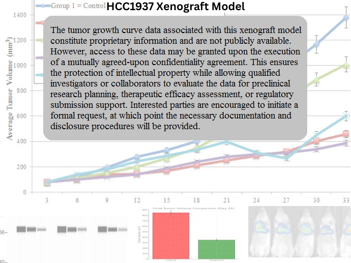
HCC1937 Xenograft Model Overview
The HCC1937 xenograft model is derived from a human triple-negative breast cancer (TNBC) and represents one of the most widely used BRCA1-mutant systems for preclinical oncology research. The parental HCC1937 cell line was established from a primary ductal carcinoma of a 24-year-old female patient and is characterized by the absence of estrogen receptor (ER), progesterone receptor (PR), and HER2 expression—hallmarks of the TNBC subtype. Critically, HCC1937 carries a homozygous 5382insC frameshift mutation in the BRCA1 gene, leading to impaired homologous recombination repair and heightened sensitivity to DNA-damaging agents.
In vivo, HCC1937 xenografts develop as moderately growing, basal-like tumors with molecular and histological characteristics representative of BRCA1-deficient TNBC. This model is highly suitable for investigating synthetic lethality strategies, DNA repair mechanisms, and PARP inhibitor response, and serves as a powerful platform for studying treatment resistance in BRCA1-mutated breast cancer.
Request a Custom Quote for HCC1937 Xenograft ModelBiological and Molecular Characteristics
HCC1937 cells display a basal-like TNBC phenotype with a truncated, nonfunctional BRCA1 protein. The loss of BRCA1 impairs homologous recombination, creating a dependency on error-prone repair pathways such as non-homologous end joining (NHEJ). This deficiency renders the cells vulnerable to DNA-damaging agents, including platinum compounds and PARP inhibitors, and positions the model as a canonical tool for exploring synthetic lethality.
The cell line expresses cytokeratin 5/6, EGFR, and vimentin, consistent with a basal epithelial and partially mesenchymal phenotype. TP53 is mutated, further exacerbating genomic instability. Activation of the PI3K/AKT pathway and expression of survivin and BCL-XL contribute to enhanced cell survival and therapy resistance. Despite BRCA1 loss, RAD51 is expressed but mislocalized, and γH2AX accumulation is elevated following genotoxic stress, indicating persistent DNA damage.
| Characteristic | HCC1937 Profile |
|---|---|
| Tumor Type | Triple-negative breast cancer (TNBC) |
| Receptor Status | ER– / PR– / HER2– |
| BRCA1 Status | Mutant (5382insC; truncated) |
| TP53 Status | Mutant |
| RAD51 Localization | Cytoplasmic (non-functional in HR) |
| Basal Markers | CK5/6+, EGFR+, Vimentin+ |
| DNA Damage Response | γH2AX+, ATM+, ATR+, Chk1/Chk2+ (dysregulated) |
| Apoptosis/Survival Regulators | BCL-XL+, Survivin+, XIAP+ |
| PI3K/AKT Signaling | Active |
| EMT Traits | Partial (vimentin+, low E-cadherin) |
This molecular landscape supports the use of HCC1937 in studies targeting DNA repair defects, PARP inhibition, and resistance mechanisms in TNBC.
In Vivo Model Development and Tumorigenicity
HCC1937 xenografts are generated by subcutaneous implantation of 5 × 10^6 to 1 × 10^7 cells in immunodeficient mice (e.g., athymic nude or NSG). Tumor take rates typically exceed 85% when cells are injected in Matrigel. Tumors become palpable within 12–16 days and reach endpoint volumes (1,200–1,400 mm³) within 28–35 days. Growth kinetics are moderately paced but highly reproducible.
Orthotopic implantation into the mammary fat pad has been used to assess tumor microenvironment interaction, ECM remodeling, and metastatic potential. While spontaneous metastasis is rare, engineered variants of HCC1937 allow for studying invasion and distant spread. The model is compatible with luciferase transduction for longitudinal tracking and response monitoring.
Xenografts maintain stable BRCA1 deficiency, facilitating long-term studies of acquired resistance, compensatory pathway activation, and clonal evolution under therapeutic pressure. They are suitable for combination therapy trials, including agents targeting PI3K, mTOR, Chk1, and immune evasion pathways.
Request a Custom Quote for HCC1937 Xenograft ModelHistopathology and Immunohistochemical Profile
Histologically, HCC1937 xenografts present as moderately cellular tumors composed of spindle to polygonal cells with enlarged nuclei, high mitotic activity, and moderate nuclear pleomorphism. The architecture is generally solid with focal necrosis and scattered inflammatory infiltrates. Stromal involvement is limited, but peritumoral fibrosis can develop in orthotopic settings.
Immunohistochemical staining reveals strong nuclear expression of Ki-67 (45–60%), cytoplasmic expression of vimentin, and membrane-localized EGFR. Tumors lack ER, PR, and HER2 staining, consistent with their TNBC phenotype. BRCA1 staining is absent or faint, and γH2AX is diffusely positive, indicating unresolved DNA damage.
Additional markers include CK5/6 and p63, confirming basal subtype identity, while BCL-XL and survivin are diffusely expressed in viable tumor zones. Phosphorylated AKT and mTOR are detectable in cytoplasmic compartments, supporting the use of this model in signal pathway inhibition studies.
Preclinical Applications and Drug Response
The HCC1937 xenograft model is the gold standard for evaluating PARP inhibitors (e.g., olaparib, talazoparib) in BRCA1-deficient TNBC. It is also widely used for assessing platinum-based therapies (e.g., cisplatin, carboplatin), which exploit DNA crosslinking and homologous recombination failure.
Due to its PI3K/AKT activation, HCC1937 is suitable for testing targeted inhibitors such as PI3K inhibitors, mTOR antagonists, and dual PI3K/mTOR agents. Combination therapy studies involving DNA damage response inhibitors (ATR, Chk1, ATM) and anti-apoptotic agents (e.g., BCL-2 family antagonists) have shown promise in overcoming resistance.
The model is also leveraged in studies of adaptive resistance mechanisms, including re-expression of BRCA1, upregulation of NHEJ components, and epigenetic reprogramming. These features make it ideal for identifying biomarkers of sensitivity and for testing rational drug combinations.
Request This Model
To request the HCC1937 xenograft model for use in studies of BRCA1-mutant TNBC, PARP inhibitor resistance, or synthetic lethality testing, please contact us using the link below. We provide subcutaneous and orthotopic implantation support, as well as customizable imaging and pharmacodynamic protocols.
Request a Custom Quote for HCC1937 Xenograft Model