HCC1187 Xenograft Model Overview
The HCC1187 xenograft model is derived from a primary ductal carcinoma of the breast from a patient with advanced-stage triple-negative breast cancer (TNBC). This cell line was established through the Hamon Cancer Center Collection (HCC) and is notable for its basal-like molecular profile, absence of hormone receptors (ER, PR), and lack of HER2 overexpression. HCC1187 exhibits features consistent with aggressive TNBC, including high proliferation, robust tumor formation in vivo, and constitutive activation of oncogenic survival pathways.
This model is widely used in the study of DNA damage repair mechanisms, PARP inhibitor responses, and synthetic lethality frameworks, particularly due to its BRCA1 deficiency. It has also been employed in drug development efforts targeting mitotic regulators and checkpoint kinases. The HCC1187 xenograft recapitulates key clinical features of basal-like TNBC and is well suited for preclinical validation of therapies targeting DNA repair vulnerabilities and chromosomal instability.
Request a Custom Quote for HCC1187 Xenograft ModelBiological and Molecular Characteristics
HCC1187 cells are ER-negative, PR-negative, and HER2-negative, placing them firmly within the triple-negative category. They express basal cytokeratins and epidermal growth factor receptor (EGFR), which is typical of the basal-like molecular subtype. The most defining molecular feature of this model is its loss of functional BRCA1, which results in defective homologous recombination repair and a strong dependency on alternative DNA repair pathways.
These characteristics make HCC1187 cells highly sensitive to PARP inhibition and other strategies that exploit replication stress and defective genome maintenance. Additional alterations include TP53 mutation and gains in chromosomal regions associated with mitotic control genes such as AURKA and PLK1.
| Characteristic | HCC1187 Profile |
|---|---|
| Tumor Type | Primary invasive ductal breast carcinoma |
| Receptor Status | ER–, PR–, HER2– (TNBC) |
| Molecular Subtype | Basal-like |
| BRCA1 Status | Deficient (non-functional) |
| TP53 Status | Mutant |
| DNA Repair Profile | Homologous recombination-deficient |
| EGFR Status | Overexpressed |
| Mitotic Regulators | AURKA and PLK1 amplification |
| Proliferation Index | High (Ki-67 > 70%) |
| Morphology | Epithelial with limited cohesion |
These features make HCC1187 ideal for mechanistic studies involving DNA damage repair, mitotic dysregulation, and cell cycle checkpoint bypass.
In Vivo Model Development and Tumorigenicity
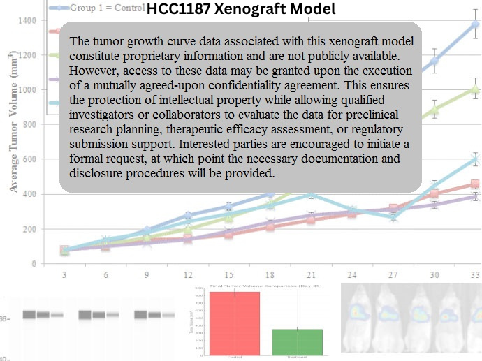
The HCC1187 xenograft model is developed by subcutaneous injection of 5–10 × 10⁶ cells into immunodeficient mice, typically NOD/SCID or athymic nude strains. The tumor take rate exceeds 90%, and nodules become palpable within 7–10 days post-implantation. The model reaches endpoint volumes of approximately 1,000–1,200 mm³ within 4–5 weeks, demonstrating consistent and aggressive in vivo growth kinetics.
Tumors exhibit a locally invasive pattern, and orthotopic implantation into the mammary fat pad can be used to mimic the native tumor microenvironment. The BRCA1-deficient status of HCC1187 allows for dynamic assessment of DNA damage accumulation, genomic instability, and therapeutic efficacy in response to genotoxic stress.
This model is well-suited to longitudinal drug efficacy studies, particularly those involving PARP inhibitors, DNA crosslinking agents, and mitotic spindle poisons. It can also be adapted for fluorescent or bioluminescent labeling to facilitate in vivo imaging.
Request a Custom Quote for HCC1187 Xenograft ModelHistopathology and Immunohistochemical Profile
HCC1187 xenografts demonstrate poorly differentiated histopathology with marked nuclear pleomorphism, high mitotic index, and minimal stromal architecture. Tumor sections show a sheet-like arrangement of tumor cells, with occasional necrotic foci and limited glandular features. The lack of differentiation is consistent with aggressive TNBC histotypes.
Immunohistochemically, tumors are negative for ER, PR, and HER2, and stain strongly for cytokeratin 5/6 and EGFR. Ki-67 labeling is frequently above 70%, confirming rapid proliferation. BRCA1 nuclear staining is absent or severely reduced, while γH2AX and phospho-RAD51 foci can be detected after DNA-damaging treatment, serving as pharmacodynamic markers for DNA repair modulation.
Phospho-AURKA and PLK1 are also detectable, highlighting mitotic dysregulation as an additional exploitable vulnerability in this model.
Preclinical Applications and Drug Response
The HCC1187 xenograft model has become a critical platform for preclinical evaluation of DNA damage response-targeted therapeutics, particularly PARP inhibitors and ATR/CHK1 inhibitors. Its BRCA1 deficiency creates a synthetic lethality framework for evaluating combination therapies that collapse replication forks or prevent cell cycle checkpoint adaptation.
The model has also been used in studies targeting mitotic progression, where its sensitivity to AURKA and PLK1 inhibitors is of translational interest. Due to its aggressive growth and genetic instability, HCC1187 is well suited for resistance evolution studies, dose-finding trials, and evaluation of multi-agent synergy in TNBC.
This model has also been included in comparative panels to evaluate TNBC subtype selectivity and has been instrumental in biomarker development for homologous recombination deficiency (HRD).
Request This Model
To incorporate the HCC1187 xenograft model into BRCA1-deficient TNBC research, PARP inhibitor testing, or mitotic checkpoint therapeutic studies, request a customized preclinical service package using the link below. Services include tumor engraftment, pharmacodynamics, DNA repair profiling, and in vivo drug screening.
Request a Custom Quote for HCC1187 Xenograft Model