GIST-T1 Xenograft Model Overview
The GIST-T1 xenograft model is derived from a human gastrointestinal stromal tumor (GIST) established from the gastric tumor of a 58-year-old Japanese male. This model harbors a homozygous activating mutation in exon 11 of the c-KIT proto-oncogene, which is the most common genetic alteration found in GISTs. GIST-T1 xenografts exhibit high fidelity to human GIST pathology, demonstrating robust and reproducible tumor growth in immunodeficient mice. Given its strong dependence on c-KIT signaling, the model is particularly suited for preclinical evaluation of tyrosine kinase inhibitors (TKIs) and downstream effector modulators, serving as a critical tool for translational research in targeted therapy for gastrointestinal stromal tumors.
Request a Custom Quote for GIST-T1 Xenograft ModelBiological and Molecular Characteristics
GIST-T1 cells possess epithelioid morphology and express classical GIST markers, including c-KIT (CD117) and DOG1. The hallmark molecular feature of this model is a constitutive activating mutation in exon 11 of the KIT gene, resulting in ligand-independent receptor activation. The cell line is negative for PDGFRA mutations, aligning it with the majority subtype of KIT-mutant GISTs. GIST-T1 is microsatellite stable (MSS) and does not harbor mutations in BRAF, KRAS, or NRAS. Downstream signaling through the PI3K/AKT and MAPK pathways is enhanced, contributing to cellular proliferation and survival. The model’s consistent receptor expression and mutation profile support its use in evaluating resistance mechanisms and combination strategies targeting multiple pathways.
| Characteristic | GIST-T1 Cell Line Profile |
|---|---|
| Tissue of Origin | Gastric GIST (primary tumor) |
| KIT Mutation | Exon 11 activating mutation |
| PDGFRA/BRAF/KRAS Status | Wild-type |
| MSI Status | Microsatellite stable (MSS) |
| Marker Expression | CD117 (c-KIT), DOG1, CD34 |
| Signaling Pathways | Activated PI3K/AKT and MAPK |
In Vivo Model Development and Tumorigenicity
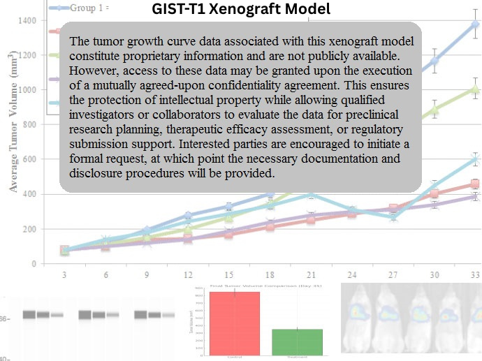
GIST-T1 xenografts are established via subcutaneous injection of cultured cells into immunodeficient mice, typically athymic nude or NOD/SCID strains. Tumor formation begins within 10–14 days post-inoculation and reaches volumes of 700–900 mm³ within 4–5 weeks. The model demonstrates high take rates and predictable growth kinetics, offering a reproducible system for pharmacodynamic and pharmacokinetic studies. Because tumor growth in GIST-T1 xenografts is driven by constitutively active c-KIT signaling, it serves as a highly responsive platform for evaluating TKIs such as imatinib, sunitinib, and regorafenib, along with emerging c-KIT antagonists and multi-kinase inhibitors.
Request a Custom Quote for GIST-T1 Xenograft ModelHistopathology and Immunohistochemical Profile
Histological analysis of GIST-T1 xenograft tumors reveals uniform epithelioid cells with eosinophilic cytoplasm, arranged in interlacing fascicles and sheets. Hematoxylin and eosin (H&E) staining shows mild nuclear pleomorphism and sparse mitotic activity, reflecting the intermediate proliferative potential of this model. Immunohistochemical staining confirms strong membranous and cytoplasmic expression of c-KIT (CD117), as well as positivity for DOG1 and CD34, aligning with clinical diagnostic features of GIST. These histologic and immunophenotypic markers confirm the fidelity of the xenograft to primary human GISTs and validate its utility for translational research targeting receptor tyrosine kinase signaling.
Preclinical Applications and Drug Response
The GIST-T1 xenograft model is widely used in evaluating the efficacy of first-, second-, and third-generation TKIs against KIT-driven GISTs. It is particularly responsive to imatinib, the frontline therapy for KIT exon 11-mutant GISTs, and is often employed to study dose optimization, resistance acquisition, and the pharmacodynamic effects of chronic TKI exposure. The model also supports preclinical testing of combination therapies that inhibit both KIT and downstream pathways such as PI3K or MEK, as well as novel agents designed to overcome imatinib resistance. Given its well-characterized mutation and reproducible in vivo behavior, GIST-T1 serves as an essential model in the development of targeted therapeutics for gastrointestinal stromal tumors.
Request This Model
To incorporate the GIST-T1 xenograft model into your GIST research program or targeted drug development pipeline, contact our scientific team to receive full model specifications and initiate a customized study strategy focused on KIT-driven gastrointestinal tumors.
Request a Custom Quote for GIST-T1 Xenograft Model