CCRF-CEM Xenograft Model Overview
The CCRF-CEM xenograft model is derived from a human T-cell acute lymphoblastic leukemia (T-ALL) line established from the peripheral blood of a 4-year-old female patient with relapsed leukemia. This model represents a classical form of T-lineage ALL, retaining key immunophenotypic and molecular features associated with immature thymic T-cell malignancies. CCRF-CEM xenografts are widely used in preclinical hematology-oncology research to investigate glucocorticoid resistance, NOTCH1 signaling, apoptosis regulation, and the efficacy of cytotoxic and targeted agents in T-ALL. Due to its reproducible engraftment and aggressive leukemic phenotype, this model supports both systemic and subcutaneous study designs and has contributed significantly to drug development pipelines targeting pediatric and relapsed ALL.
Request a Custom Quote for CCRF-CEM Xenograft ModelBiological and Molecular Characteristics
CCRF-CEM cells exhibit a round, non-adherent suspension morphology and express typical markers of early T-cell differentiation, including CD3, CD7, CD5, and CD1a. The model lacks B-lineage markers (CD19, CD20) and shows strong nuclear expression of TdT (terminal deoxynucleotidyl transferase), indicative of an immature lymphoblastic phenotype. CCRF-CEM cells are wild-type for p53 but possess abnormalities in glucocorticoid receptor signaling, particularly NR3C1 downregulation, which contributes to intrinsic steroid resistance. The cell line exhibits activation of NOTCH1 and PI3K/AKT signaling pathways and has demonstrated sensitivity to drugs modulating apoptosis (e.g., BCL-2 inhibitors). Chromosomal analysis reveals a hyperdiploid karyotype with structural rearrangements involving chromosomes 7, 9, and 14.
| Characteristic | CCRF-CEM Cell Line Profile |
|---|---|
| Disease Origin | T-cell acute lymphoblastic leukemia (T-ALL) |
| Immunophenotype | CD3⁺, CD7⁺, CD5⁺, CD1a⁺, TdT⁺ |
| p53 Status | Wild-type |
| Glucocorticoid Response | Partial resistance (↓NR3C1 expression) |
| NOTCH1 Status | Activated pathway |
| Key Pathways | PI3K/AKT, apoptosis, glucocorticoid signaling |
In Vivo Model Development and Tumorigenicity
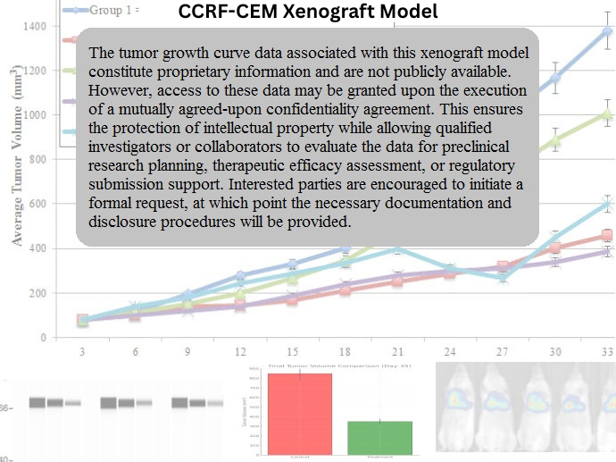
CCRF-CEM xenografts are established in immunodeficient mice, including NOD/SCID or NSG strains, using either subcutaneous or intravenous injection. Subcutaneous models allow for quantifiable tumor volume endpoints, while systemic (disseminated) models more accurately replicate the clinical pathology of T-ALL, including infiltration of the spleen, bone marrow, and central nervous system. In subcutaneous studies, tumors typically reach volumes of 700–900 mm³ within 3–5 weeks and maintain consistent take rates. The CCRF-CEM model is well suited for evaluating drug response kinetics, testing novel immunotherapies, and investigating leukemic persistence under therapeutic pressure, particularly in glucocorticoid-refractory T-ALL settings.
Request a Custom Quote for CCRF-CEM Xenograft ModelHistopathology and Immunohistochemical Profile
Histological analysis of CCRF-CEM xenografts shows diffuse sheets of small-to-medium lymphoblasts with scant cytoplasm, high nuclear-to-cytoplasmic ratio, fine chromatin, and inconspicuous nucleoli. H&E staining reveals a uniform blast population without significant necrosis or stromal involvement in early-stage tumors. Immunohistochemically, xenografts express strong nuclear TdT, surface CD3 and CD7, and cytoplasmic CD5, in keeping with their immature T-cell lineage. Phosphorylated AKT and BCL-2 expression are also observed, supporting their utility in signal transduction and apoptosis-targeted studies. CNS-infiltrating disease in systemic models can be evaluated through IHC staining of leptomeningeal tissues for T-cell markers and TdT.
Preclinical Applications and Drug Response
The CCRF-CEM xenograft model has been extensively applied in studies evaluating chemotherapeutic regimens used in pediatric T-ALL, including vincristine, dexamethasone, and L-asparaginase. Its partial resistance to glucocorticoids makes it particularly valuable for studying steroid-refractory disease mechanisms. Additionally, the model supports testing of targeted agents such as BCL-2 inhibitors (e.g., venetoclax), NOTCH pathway antagonists, and PI3K/AKT modulators. CCRF-CEM xenografts have also been used to validate T-cell immunotherapies and bi-specific antibodies in preclinical systems. Their robust engraftment and disease recapitulation make them a foundational model in translational T-ALL research.
Request This Model
To incorporate the CCRF-CEM xenograft model into your preclinical T-ALL investigations or therapeutic development programs, contact our scientific team for comprehensive model specifications and tailored support in designing both subcutaneous and disseminated leukemia studies.
Request a Custom Quote for CCRF-CEM Xenograft Model