Calu-1 Xenograft Model Overview
The Calu-1 xenograft model is established from a human non-small cell lung carcinoma (NSCLC) squamous cell line originally derived from a male patient with epidermoid carcinoma of the lung. This model is frequently used in preclinical oncology to evaluate therapeutic responses in KRAS-mutant, EGFR-wild-type NSCLC, making it a valuable counterpart to adenocarcinoma-based xenograft systems. The Calu-1 model is particularly suited for studies of apoptosis, cell cycle dysregulation, and pathway-specific inhibition in squamous cell carcinomas, and is commonly used in both monotherapy and combinatorial drug efficacy evaluations involving chemotherapy and small molecule inhibitors.
Request a Custom Quote for Calu‑1 Xenograft ModelBiological and Molecular Characteristics
Calu-1 cells are characterized by a robust epithelial phenotype and possess several defining molecular features, including a homozygous deletion of the TP53 gene and a KRAS G12C activating mutation. This profile positions the model as a representative of squamous NSCLC subtypes with mutant RAS pathway activation and defective DNA damage response. The line expresses cytokeratins (e.g., CK5/6) and basal squamous markers, while lacking neuroendocrine or glandular differentiation features. EGFR is expressed at moderate levels without activating mutations, making Calu-1 ideal for testing therapies that target downstream elements of the RAS/RAF/MEK/ERK cascade or synthetic lethality approaches in p53-null tumors.
| Characteristic | Calu-1 Cell Line Profile |
|---|---|
| Cancer Type | Non-small cell lung carcinoma (squamous cell) |
| KRAS Status | Mutant (G12C) |
| TP53 Status | Homozygous deletion |
| EGFR Status | Wild-type |
| Key Pathways | RAS/RAF/MEK, PI3K/AKT |
| Marker Expression | Cytokeratin⁺, CK5/6⁺, p63⁺ |
In Vivo Model Development and Tumorigenicity
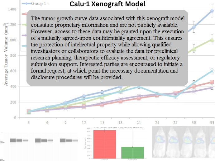
Calu-1 xenografts are reliably formed through subcutaneous injection of cultured cells into immunodeficient murine hosts such as nude or NOD/SCID mice. Tumors exhibit moderate to rapid growth kinetics, with palpable masses forming within 7–10 days and reaching volumes of 700–900 mm³ over approximately 4–5 weeks. The model demonstrates consistent tumor take rates and reproducibility across cohorts, making it suitable for longitudinal therapeutic trials. Given its squamous origin, Calu-1 may also be adapted to orthotopic or lung-specific implantation strategies to more closely replicate tissue architecture and drug distribution profiles in vivo.
Request a Custom Quote for Calu‑1 Xenograft ModelHistopathology and Immunohistochemical Profile
Calu-1 xenografts display histologic patterns consistent with poorly to moderately differentiated squamous cell carcinoma. Tumor sections reveal nests and sheets of polygonal cells with eosinophilic cytoplasm, intercellular bridges, and high nuclear-to-cytoplasmic ratios. Keratinization is minimal, reflecting an undifferentiated squamous phenotype. Immunohistochemical analyses demonstrate positivity for cytokeratin 5/6, p63, and pancytokeratin, while TTF-1 and surfactant markers are absent, confirming non-adenocarcinoma lineage. Ki-67 labeling indices are high, correlating with rapid proliferation. p53 staining is absent due to genomic deletion, while phosphorylated ERK and AKT highlight constitutive pathway activation downstream of KRAS mutation.
Preclinical Applications and Drug Response
The Calu-1 xenograft model is an established system for evaluating KRAS G12C inhibitors, p53 synthetic lethality agents, and MEK/ERK pathway modulators. Its use has expanded in immuno-oncology and combination therapy studies, particularly in settings where EGFR is not a viable target. The model’s p53-null status allows mechanistic dissection of cell cycle checkpoint inhibitors, DNA damage response modulators, and apoptosis-inducing agents. Calu-1 has also demonstrated utility in screening chemotherapeutics such as cisplatin and docetaxel, and serves as a comparator for adenocarcinoma xenografts in subtype-specific drug response evaluations.
Request This Model
To incorporate the Calu-1 xenograft model into your squamous NSCLC research or drug development workflow, contact our team to access validated model protocols, tumor growth data, and expert guidance tailored to KRAS-mutant, p53-deficient lung cancer studies.
Request a Custom Quote for Calu‑1 Xenograft Model