A204 Xenograft Model Overview
The A204 xenograft model is derived from a human rhabdomyosarcoma (RMS) cell line, A204, established from a metastatic lesion in the pelvis of a patient with alveolar rhabdomyosarcoma, a subtype of RMS. Rhabdomyosarcoma is the most common soft tissue sarcoma in children and adolescents, and it is characterized by aggressive growth, frequent metastasis, and resistance to chemotherapy. The A204 xenograft model is highly valuable for preclinical research as it replicates key features of rhabdomyosarcoma, including tumor progression, metastatic spread, and chemoresistance. This model is widely used to evaluate novel therapeutic strategies, including chemotherapy, targeted therapies, and immunotherapies, aimed at improving treatment outcomes for RMS patients.
Request a Custom Quote for A204 Xenograft ModelBiological and Molecular Characteristics
A204 cells are characterized by their mesenchymal origin and ability to form skeletal muscle-like structures. These cells exhibit strong expression of muscle markers, including desmin, myogenin, and myosin, which are indicative of their rhabdomyosarcoma origin. The A204 model also expresses the PAX3-FOXO1 fusion gene, a key driver of alveolar rhabdomyosarcoma, which contributes to the tumor’s aggressive nature and metastatic potential. Additionally, A204 cells exhibit dysregulated signaling in pathways such as PI3K/AKT and MAPK/ERK, which contribute to cell survival, proliferation, and invasion. These molecular features make the A204 xenograft model highly relevant for studying the biology of rhabdomyosarcoma and for testing therapies that target these pathways.
| Marker | Expression Level | Function |
|---|---|---|
| Desmin | High | Muscle marker associated with rhabdomyosarcoma |
| Myogenin | High | Muscle differentiation marker |
| PAX3-FOXO1 Fusion | Present | Oncogene fusion in alveolar rhabdomyosarcoma |
| PI3K/AKT pathway | Dysregulated | Promotes cell survival and proliferation |
In Vivo Model Development and Tumorigenicity
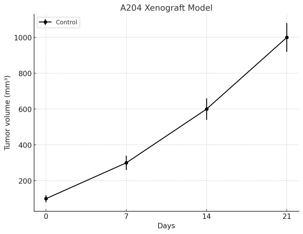
The A204 xenograft model is typically established by implanting A204 cells into immunocompromised mice, such as NOD/SCID or NSG mice, which lack functional T and B cells. Once implanted, the cells form rapidly growing tumors that closely resemble human rhabdomyosarcoma in terms of histology, cellular structure, and metastatic behavior. The tumors exhibit high cellularity, abundant mitotic activity, and significant vascularization due to the angiogenic properties of the tumor. Given its ability to replicate key features of RMS, including its aggressive growth and metastasis to distant organs, the A204 xenograft model is particularly useful for evaluating chemotherapy agents, including doxorubicin and ifosfamide, which are commonly used in RMS treatment regimens.
In addition to subcutaneous implantation, orthotopic models of A204 can be established by implanting the cells into the skeletal muscle of immunocompromised mice. This orthotopic model more closely mimics the natural site of tumor growth and allows for the study of local invasion, metastasis, and the effects of treatment on tumor progression. A204 cells are capable of metastasizing to distant organs, particularly the lungs and lymph nodes, making this model ideal for studying metastatic disease and evaluating therapies aimed at preventing or treating metastasis in rhabdomyosarcoma.
Request a Custom Quote for A204 Xenograft ModelHistopathology and Immunohistochemical Profile
Histopathological examination of A204 xenografts reveals the characteristic features of rhabdomyosarcoma, including round to polygonal tumor cells, frequent mitotic activity, and abundant cytoplasm. The tumors also exhibit areas of necrosis, indicative of rapid tumor growth. Immunohistochemical staining of A204 xenografts shows strong expression of muscle-specific markers such as desmin and myogenin, confirming the skeletal muscle origin of the tumor. Additionally, the tumors show the presence of the PAX3-FOXO1 fusion gene, which is detected using specific probes and plays a key role in the pathogenesis of alveolar rhabdomyosarcoma. High levels of phosphorylated AKT are observed in the tumors, indicating activation of the PI3K/AKT pathway, which promotes cell survival and resistance to therapy. CD31 staining reveals significant angiogenesis, reflecting the high vascularity of the tumors, which is critical for sustaining tumor growth.
Preclinical Applications and Drug Response
The A204 xenograft model is widely used to evaluate the efficacy of various chemotherapy agents for rhabdomyosarcoma, including doxorubicin, ifosfamide, and other standard treatments. The model’s ability to develop resistance to chemotherapy over time makes it particularly useful for studying the mechanisms of drug resistance and for testing new agents aimed at overcoming these challenges. Given the presence of the PAX3-FOXO1 fusion gene and the dysregulated signaling in the PI3K/AKT and MAPK pathways, the A204 xenograft model is also highly valuable for evaluating targeted therapies that aim to inhibit these pathways.
In addition to chemotherapy and targeted therapies, the A204 xenograft model is increasingly used to assess the potential of immunotherapies, including immune checkpoint inhibitors and monoclonal antibodies targeting tumor-specific antigens. The model’s ability to replicate key features of rhabdomyosarcoma, including its aggressive growth, bone and soft tissue involvement, and metastatic potential, makes it an ideal platform for studying new treatment strategies. Furthermore, the A204 model is useful for investigating combination therapies, where chemotherapy is combined with novel immunotherapies or targeted agents to improve treatment efficacy and prevent drug resistance.
Request This Model
To request the A204 xenograft model for your preclinical studies, please use the form below. A customized quote and additional model specifications will be provided upon inquiry.
Request a Custom Quote for A204 Xenograft Model