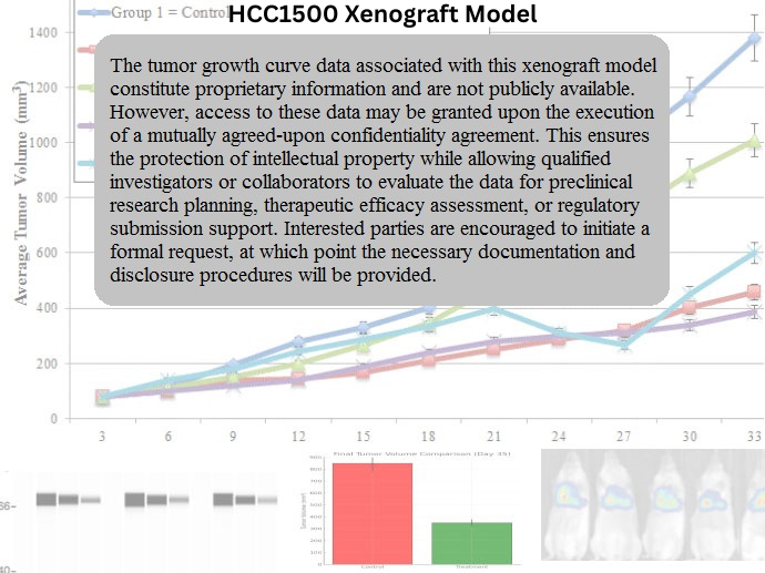
HCC1500 Xenograft Model Overview
The HCC1500 xenograft model originates from a human primary breast tumor classified as invasive ductal carcinoma. The cell line was derived from a 60-year-old female patient and is part of the Hamon Cancer Center Collection (HCC). HCC1500 cells are estrogen receptor-positive (ER⁺), progesterone receptor-negative (PR⁻), and HER2-negative (HER2⁻), making this model representative of luminal-type breast cancers with partial hormone dependency.
This model is particularly relevant for studying resistance to anti-estrogen therapies, evaluating ER signaling modulation, and profiling the molecular responses of luminal B-like tumors. It is a reliable system for assessing single-agent or combination therapies involving ER antagonists, CDK4/6 inhibitors, and PI3K/AKT/mTOR pathway modulators. The HCC1500 xenograft model combines moderate tumorigenicity with clinically relevant expression patterns, making it a valuable tool in translational breast cancer research.
Request a Custom Quote for HCC1500 Xenograft ModelBiological and Molecular Characteristics
HCC1500 exhibits a luminal epithelial phenotype with ER expression and moderate proliferative activity. Although progesterone receptor expression is absent, the ER-positive, HER2-negative status places the model in the luminal B category. This subtype is characterized by a higher proliferation index compared to luminal A tumors and often displays suboptimal responses to endocrine monotherapy, particularly in the absence of PR signaling.
The cell line demonstrates intact BRCA1/2 function, wild-type TP53, and modest activation of PI3K/AKT signaling. The absence of HER2 amplification and low baseline EMT marker expression underscore its epithelial stability and hormone-driven proliferation.
| Characteristic | HCC1500 Profile |
|---|---|
| Tumor Type | Invasive ductal breast carcinoma |
| Receptor Status | ER⁺, PR⁻, HER2⁻ |
| Molecular Subtype | Luminal B |
| Estrogen Dependency | Moderate |
| HER2 Status | IHC 1+ / FISH-negative |
| TP53 Status | Wild-type |
| PI3K/AKT Pathway | Modestly active |
| EMT Markers | Low |
| Proliferation Index | Moderate (Ki-67 ~30–40%) |
| Differentiation Status | Epithelial, well organized |
The absence of PR expression makes this model well suited for investigating incomplete endocrine responses and PR-independent ER signaling.
In Vivo Model Development and Tumorigenicity
HCC1500 xenografts are typically established by injecting 5–10 × 10⁶ cells subcutaneously into immunodeficient mice, such as athymic nude or NOD/SCID strains. Estrogen supplementation (via subcutaneous 17β-estradiol pellets) enhances tumor take and growth, though some tumors can establish without hormonal support, reflecting partial estrogen dependence.
Tumor take rates are approximately 75–85% with estrogen supplementation, and xenografts reach volumes of 1,000–1,200 mm³ over a 6–8 week period. Growth kinetics are consistent, with limited necrosis and a low rate of spontaneous regression. Orthotopic implantation into the mammary fat pad can further enhance the fidelity of tumor–host interaction modeling.
This model supports molecular imaging, longitudinal therapy tracking, and ex vivo tissue analysis for pharmacodynamic readouts.
Request a Custom Quote for HCC1500 Xenograft ModelHistopathology and Immunohistochemical Profile
Histologically, HCC1500 xenografts are composed of moderately differentiated epithelial cells arranged in glandular patterns with minimal stromal infiltration. Nuclear morphology is uniform, with occasional mitotic figures and limited necrotic foci. The overall architecture reflects a low-to-moderate grade carcinoma consistent with luminal B biology.
Immunohistochemical analysis confirms strong nuclear ER staining, negative PR expression, and HER2 scores of 0 to 1+. E-cadherin is present, maintaining epithelial integrity. The Ki-67 index ranges between 30% and 40%, supporting moderate proliferative potential. In response to endocrine therapy, tumors demonstrate downregulation of ER and decreased Ki-67, which serve as reliable pharmacodynamic biomarkers.
Additional markers such as BCL2 and cyclin D1 are variably expressed and can be modulated in response to drug treatment, enabling mechanistic exploration of apoptotic resistance and cell cycle arrest.
Preclinical Applications and Drug Response
The HCC1500 xenograft model is particularly suited for evaluating endocrine therapies such as tamoxifen, fulvestrant, and aromatase inhibitors, especially in PR-negative contexts. It is also appropriate for studies of endocrine resistance mechanisms and the role of co-targeting signaling pathways such as PI3K/AKT or CDK4/6.
This model supports dual therapy testing with ER antagonists and small molecule inhibitors of the mTOR or MAPK pathways. The absence of HER2 overexpression makes it ideal for studies isolating the effects of ER modulation without HER2-driven interference. Furthermore, its partial hormone dependence permits the evaluation of next-generation SERDs and hybrid therapies designed to address incomplete endocrine responses.
The HCC1500 model provides a robust luminal B system for exploring predictive biomarkers, treatment adaptation, and durable tumor control strategies.
Request This Model
To utilize the HCC1500 xenograft model in your hormone-responsive breast cancer study or targeted drug development program, request a custom xenograft services package below. Offerings include tumor establishment, hormone modulation, longitudinal therapy trials, and molecular endpoint analysis.
Request a Custom Quote for HCC1500 Xenograft Model