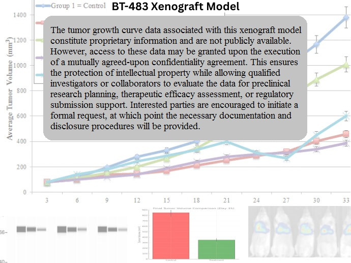
BT-483 Xenograft Model Overview
The BT-483 xenograft model is derived from a human breast carcinoma exhibiting luminal characteristics. This cell line originates from a ductal carcinoma of the breast and is estrogen receptor-positive (ER⁺), progesterone receptor-positive (PR⁺), and HER2-low, representing a clinically relevant subtype of hormone-driven breast cancer. BT-483 is well-suited for evaluating anti-estrogen therapies, combination hormone-targeted regimens, and resistance mechanisms to endocrine therapies.
Unlike triple-negative or HER2-amplified models, BT-483 exhibits slower tumor growth and is dependent on hormonal stimulation for robust in vivo proliferation. This model offers high utility in studies of estrogen responsiveness, receptor modulation, and the development of resistance to selective estrogen receptor modulators (SERMs) and aromatase inhibitors.
Request a Custom Quote for BT-483 Xenograft ModelBiological and Molecular Characteristics
BT-483 cells express both estrogen and progesterone receptors and require estrogenic signaling for optimal proliferation. The cell line demonstrates low-level HER2 expression but is not HER2-amplified. Its molecular profile is representative of the luminal B subtype, which is associated with higher proliferation than luminal A and a greater likelihood of endocrine therapy resistance.
BT-483 cells also exhibit intact p53 function, modest PI3K/AKT pathway activation, and low basal levels of epithelial-mesenchymal transition (EMT) markers. These traits support its application in studying the molecular underpinnings of ER-driven growth and response modulation by hormone-based therapies.
| Characteristic | BT-483 Profile |
|---|---|
| Tumor Type | Invasive ductal breast carcinoma |
| Receptor Status | ER⁺, PR⁺, HER2⁻ (low expression, not amplified) |
| Molecular Subtype | Luminal B |
| Estrogen Dependency | High |
| HER2 Status | HER2 IHC 1+/2⁺; FISH-negative |
| PI3K/AKT Pathway | Modestly active |
| TP53 Status | Wild-type |
| EMT Markers | Low |
| Proliferation Index | Moderate (Ki-67 ~25–35%) |
| Differentiation Status | Well-differentiated epithelial |
These characteristics make BT-483 an ideal model for evaluating classical and next-generation endocrine therapies in ER⁺ tumors.
In Vivo Model Development and Tumorigenicity
BT-483 xenografts are developed by subcutaneous implantation of 5–10 × 10⁶ cells into immunodeficient mice, typically athymic nude or NSG strains. Due to the hormone-responsive nature of the cell line, estrogen supplementation (e.g., via subcutaneously implanted 17β-estradiol pellets) is required to achieve consistent tumor take and growth.
Tumor formation begins within 14–18 days post-injection, and volumes of 1,000–1,200 mm³ are typically reached over a 6–8 week period. The tumor take rate is approximately 70–80% with estrogen supplementation, and tumors display uniform growth patterns without extensive necrosis or hemorrhage.
The model is compatible with orthotopic implantation into the mammary fat pad, providing enhanced microenvironmental relevance. It is also amenable to longitudinal pharmacodynamic monitoring and molecular endpoint analysis of receptor expression, cell proliferation, and apoptosis induction.
Request a Custom Quote for BT-483 Xenograft ModelHistopathology and Immunohistochemical Profile
Histologically, BT-483 xenografts are composed of well-differentiated glandular structures with a relatively low nuclear grade and organized epithelial architecture. The tumor morphology reflects the underlying luminal nature of the cell line and closely mirrors hormone receptor-positive human tumors.
Immunohistochemical analysis reveals strong nuclear ER and PR staining, with low to intermediate membranous HER2 expression (1+ to 2+, without gene amplification). Ki-67 staining is moderate, corresponding to a proliferation rate of 25–35%. E-cadherin staining is retained, consistent with epithelial integrity, and phospho-AKT levels are modest, indicating baseline survival signaling that can be modulated therapeutically.
Upon anti-estrogen treatment, reductions in ER expression and Ki-67 are detectable, providing quantitative biomarkers of drug response.
Preclinical Applications and Drug Response
The BT-483 xenograft model is highly suitable for the preclinical evaluation of endocrine therapies including tamoxifen, fulvestrant, aromatase inhibitors (e.g., letrozole), and selective estrogen receptor degraders (SERDs). It provides a robust platform for assessing response dynamics, treatment duration effects, and the emergence of acquired resistance.
In combination studies, BT-483 supports co-treatment with PI3K inhibitors, CDK4/6 inhibitors, and mTOR inhibitors—especially in modeling luminal B breast cancers with moderate proliferative activity. Its intact TP53 status and differentiated phenotype also allow for mechanistic studies of hormone-receptor signaling, transcriptional reprogramming, and tumor dormancy.
Because BT-483 does not overexpress HER2, it provides a clean system for isolating the effects of hormone signaling without confounding HER2-driven oncogenic pathways.
Request This Model
To incorporate the BT-483 xenograft model into your hormone receptor-positive breast cancer study or preclinical endocrine therapy screening, request a custom xenograft services package at the link below. Services include tumor engraftment, estrogen supplementation, longitudinal therapy response tracking, and molecular endpoint analysis.
Request a Custom Quote for BT-483 Xenograft Model