MT-3 Xenograft Model Overview
The MT-3 xenograft model originates from a metastatic human breast adenocarcinoma and is characterized by its overexpression of the HER2/neu proto-oncogene. It serves as a robust preclinical system for modeling HER2-positive breast cancer, especially for evaluating therapeutic agents targeting HER2 and downstream signal transduction pathways. The MT-3 cell line was derived from pleural effusion fluid of a patient with metastatic disease, and it maintains a highly tumorigenic phenotype when transplanted into immunocompromised mice.
The MT-3 model exhibits aggressive growth, consistent HER2 amplification, and high mitotic activity, making it well suited for evaluating pharmacodynamic responses to monoclonal antibodies, small molecule kinase inhibitors, and combination regimens aimed at overcoming primary or acquired resistance to HER2-targeted therapies. Its strong HER2 dependency allows for clear evaluation of on-target drug activity and therapeutic modulation of HER2-driven signaling.
Request a Custom Quote for MT-3 Xenograft ModelBiological and Molecular Characteristics
MT-3 cells demonstrate high-level HER2 overexpression driven by ERBB2 gene amplification, classifying them within the HER2-enriched molecular subtype of breast cancer. The model lacks estrogen receptor (ER) and progesterone receptor (PR) expression, which allows for isolation of HER2-related oncogenic behavior without hormonal crosstalk. Downstream signaling pathways such as MAPK/ERK and PI3K/AKT are constitutively active, and MT-3 cells display heightened sensitivity to HER2 blockade under baseline conditions.
The cells exhibit an epithelial morphology, and their surface profile is defined by robust HER2 membrane localization and minimal expression of basal or mesenchymal markers. No common hotspot mutations in PIK3CA or TP53 have been reported, allowing drug responses to be attributed largely to HER2-driven signaling dynamics.
| Characteristic | MT-3 Profile |
|---|---|
| Tumor Type | Human breast adenocarcinoma (HER2+) |
| ER / PR Status | ER–, PR– |
| HER2 Status | Overexpressed (ERBB2 amplification) |
| Molecular Subtype | HER2-enriched |
| PI3K/AKT Pathway | Activated |
| MAPK/ERK Pathway | Activated |
| PTEN Status | Intact |
| p53 Status | Wild-type |
| Growth Factor Dependence | HER2-dependent; ligand-independent activation |
| Doubling Time | ~24 hours (in vitro); rapid in vivo growth |
This molecular profile positions MT-3 as a reliable model for testing HER2-specific inhibitors, resistance mechanisms, and targeted signaling disruptions.
In Vivo Model Development and Tumorigenicity
MT-3 xenografts are established via subcutaneous injection of 5 × 10⁶ to 1 × 10⁷ cells into the flanks of athymic nude or NOD/SCID mice. Tumor formation occurs rapidly, with nodules detectable as early as day 5 post-injection and tumors reaching 1,000–1,200 mm³ within 3–4 weeks. Take rates routinely exceed 95%, and in vivo HER2 expression is stably retained across passages.
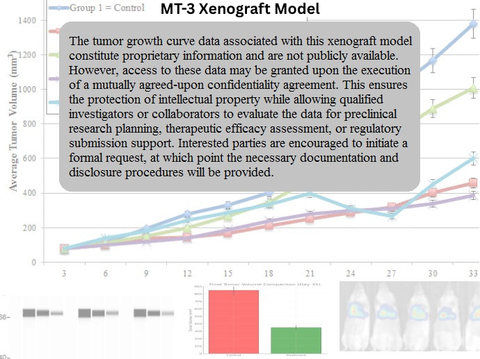
This model is also amenable to orthotopic implantation into the mammary fat pad, which enhances microenvironmental fidelity for drug distribution, stromal interactions, and localized angiogenesis studies. MT-3 is frequently used in longitudinal pharmacology studies, given its consistent and aggressive growth curve, allowing precise scheduling of multi-dose regimens and tumor response monitoring.
Fluorescent or luciferase tagging of MT-3 cells enables non-invasive imaging of tumor progression and therapeutic efficacy in live animal models. The model’s reproducibility and HER2 dependence support its use as a control or comparator in panels of HER2-driven breast cancer xenografts.
Request a Custom Quote for MT-3 Xenograft ModelHistopathology and Immunohistochemical Profile
Histologically, MT-3 xenografts resemble poorly differentiated invasive ductal carcinomas, with dense cellularity, high nuclear-to-cytoplasmic ratio, and frequent mitotic figures. Tumors lack glandular differentiation and often present with central necrosis due to rapid expansion and insufficient perfusion. A fibrotic stromal component is sometimes observed, especially in long-term passages.
Immunohistochemistry reveals intense membranous HER2 staining (3+), consistent with strong ERBB2 amplification. ER and PR staining are negative. Ki-67 staining is typically high (60–80%), reflecting the rapid proliferation rate. Phospho-AKT and phospho-ERK staining are both strongly positive, affirming downstream signaling activation.
Markers of apoptosis such as cleaved PARP and caspase-3 are inducible following HER2 blockade, supporting the use of this model for therapeutic mechanism-of-action studies. No EMT features are observed, and the tumor maintains an epithelial immunophenotype (e.g., E-cadherin positive, vimentin negative).
Preclinical Applications and Drug Response
The MT-3 xenograft model is widely used in the development of HER2-targeted therapies, including monoclonal antibodies (trastuzumab, pertuzumab), tyrosine kinase inhibitors (lapatinib, neratinib), and antibody-drug conjugates (T-DM1, trastuzumab deruxtecan). Its high HER2 dependency makes it ideal for benchmarking drug potency and for studying pharmacodynamic endpoints such as HER2 internalization, ubiquitination, and signaling pathway inhibition.
This model is also well suited for examining drug resistance mechanisms including HER2 truncation (p95HER2), compensatory receptor signaling (e.g., EGFR or HER3), and downstream reactivation of PI3K/AKT or mTOR pathways. It has been employed in studies evaluating dual pathway blockade (HER2 + mTOR, HER2 + CDK4/6) and in identifying biomarkers predictive of response or relapse in HER2+ disease.
Due to its epithelial stability and lack of hormone receptor expression, MT-3 is an excellent tool for isolating HER2-specific effects without hormonal confounding, and it is often included in preclinical screening panels for FDA IND-enabling studies.
Request This Model
To utilize the MT-3 xenograft model in HER2-targeted drug development, combination therapy design, or mechanistic signaling research, request a customized service package below. Services include model engraftment, longitudinal imaging, histopathology, and multi-agent drug screening.
Request a Custom Quote for MT-3 Xenograft Model