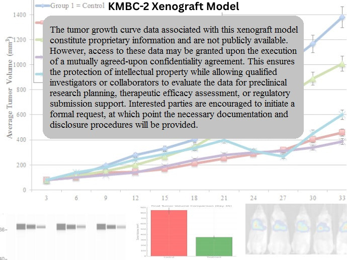
KMBC-2 Xenograft Model Overview
The KMBC-2 xenograft model is derived from a human bladder carcinoma cell line originally isolated from a moderately differentiated transitional cell carcinoma. Although less commonly used than other bladder cancer lines, KMBC-2 provides a unique molecular and biological profile that bridges features of non–muscle-invasive and early muscle-invasive urothelial carcinoma. The cell line demonstrates a relatively moderate growth rate in vivo, combined with defined epithelial morphology, partial differentiation, and a functional DNA damage response axis, allowing it to serve as a biologically distinct platform for preclinical bladder cancer studies.
In xenograft applications, KMBC-2 tumors grow with consistent latency and reach study endpoints in a predictable manner. The model is particularly well suited for investigating EGFR-driven proliferation, intermediate-grade therapeutic resistance, and pathways that govern epithelial stability and survival. KMBC-2 is increasingly recognized for its utility in studies exploring drug-induced differentiation, modulation of proliferative signaling, and histone-targeted epigenetic therapies, especially where a moderate aggressiveness model is desired.
Request a Custom Quote for KMBC-2 Xenograft ModelBiological and Molecular Characteristics
KMBC-2 cells possess a moderately epithelial phenotype, retaining E-cadherin, cytokeratin 18, and CK7 expression while lacking full urothelial terminal differentiation markers such as uroplakin II and CK20. The cells harbor wild-type TP53 and RB1, distinguishing them from more genomically unstable or p53-deficient bladder cancer models. KMBC-2 also expresses moderate levels of EGFR and HER2/ERBB2, making it suitable for studies targeting receptor tyrosine kinases and their downstream signaling cascades.
The cell line lacks activating mutations in FGFR3, but displays mild upregulation of PI3K/AKT and MAPK/ERK signaling, primarily under stress conditions. It demonstrates low-to-moderate expression of matrix metalloproteinases, indicating limited invasive potential. Notably, KMBC-2 has intact apoptotic signaling pathways, and its response to DNA damage is both dose- and time-dependent.
| Characteristic | KMBC-2 Profile |
|---|---|
| Origin | Human transitional cell carcinoma, bladder |
| TP53 Status | Wild-type |
| RB1 Status | Wild-type |
| EGFR Expression | Moderate |
| HER2/ERBB2 Expression | Moderate |
| Differentiation Markers | CK7+, CK18+, E-cadherin+ |
| Terminal Urothelial Markers | Uroplakin II-, CK20- |
| MMP Expression | Low-to-moderate (MMP-2+, MMP-9 low) |
| Apoptotic Pathway | Intact (caspase-3/7 responsive) |
| Growth Factor Signaling | Basal PI3K/AKT and MAPK/ERK activity |
This unique combination of epithelial integrity, moderate proliferative signaling, and functional apoptosis places KMBC-2 in a favorable position for studying reversible differentiation, drug responsiveness, and regulatory feedback loops in early-stage urothelial carcinoma.
In Vivo Model Development and Tumorigenicity
KMBC-2 cells form tumors efficiently in immunodeficient mice, with take rates typically exceeding 85% in athymic nude or NOD/SCID hosts. Subcutaneous implantation of 5 × 10^6 to 1 × 10^7 cells mixed in Matrigel results in palpable tumor formation within 12–15 days. Tumor growth follows a steady, reproducible trajectory, with final volumes of 1,200–1,400 mm³ typically achieved by 28–35 days post-injection.
Tumors exhibit compact, cohesive masses without overt invasiveness or necrosis, allowing for straightforward caliper-based volume monitoring and tissue sampling. Orthotopic implantation into the bladder wall has been piloted in advanced studies, with localized growth but minimal stromal penetration observed. Due to their slower kinetics compared to mesenchymal or basal-subtype bladder models, KMBC-2 xenografts are ideal for longer-duration therapeutic windows or delayed onset treatment regimens.
The model is suitable for pharmacokinetic, pharmacodynamic, and histological assessment and supports bioluminescent imaging when appropriately transduced. The predictable tumor progression enables consistent baseline comparisons and longitudinal evaluation of therapeutic agents.
Request a Custom Quote for KMBC-2 Xenograft ModelHistopathology and Immunohistochemical Profile
KMBC-2 xenografts demonstrate histopathologic features of moderately differentiated transitional cell carcinoma, with polygonal epithelial cells arranged in nests and cords. Nuclei are round to oval with visible nucleoli, and mitotic activity is moderate. Tumors exhibit low stromal content and a well-defined periphery, with minimal necrosis under standard growth conditions.
Immunohistochemically, KMBC-2 tumors are positive for E-cadherin, CK18, and CK7, affirming epithelial origin. EGFR and HER2 show membranous staining at moderate intensity, while Ki-67 index typically ranges from 35–50%, indicating a steady proliferative rate. Vimentin and N-cadherin are absent, confirming the lack of mesenchymal differentiation.
Staining for phospho-AKT and phospho-ERK1/2 is evident in cytoplasmic compartments, particularly at the leading edge of expanding tumor margins. Caspase-3 activation can be detected in response to treatment with DNA-damaging agents, confirming the presence of functional intrinsic apoptotic machinery. Overall, KMBC-2 tumors preserve epithelial architecture and histological stability across study durations.
Preclinical Applications and Drug Response
The KMBC-2 xenograft model is suited for evaluating EGFR/HER2-targeted therapies, epigenetic modulators, and agents that engage functional apoptosis pathways. Because the model retains wild-type TP53 and RB1, it serves as a control or comparator system for drugs that depend on p53-mediated apoptosis or intact checkpoint activation. The moderate expression of EGFR and HER2 allows for controlled sensitivity to TKIs such as erlotinib and lapatinib, though complete tumor regression is rarely observed without combination therapy.
KMBC-2 has demonstrated responsiveness to histone deacetylase inhibitors (HDACi), with evidence of upregulated pro-apoptotic gene expression and altered differentiation marker profiles. It also serves as a valuable system for testing drugs that modulate chromatin structure, transcriptional activation, or reversible differentiation processes. The presence of functional apoptotic machinery makes it appropriate for studies involving BCL-2 antagonists, TRAIL agonists, or DNA-alkylating agents.
Although not a highly invasive model, KMBC-2 is useful in evaluating early-stage tumor progression, epithelial maintenance, and pharmacologic reversal of proliferative signaling. It is especially well suited for experiments seeking to maintain or restore urothelial differentiation while suppressing growth via targeted therapeutic interventions.
Request This Model
To request the KMBC-2 xenograft model or explore integration into a drug screening platform focused on apoptosis, receptor inhibition, or differentiation therapy, please contact our team using the quote request form below. We support fully customized model implementation strategies and detailed technical consultation.
Request a Custom Quote for KMBC-2 Xenograft Model