HT1197 Xenograft Model Overview
The HT1197 xenograft model is derived from a human bladder carcinoma cell line established from a high-grade transitional cell carcinoma of the urinary bladder. The HT1197 cell line was isolated from a female patient with invasive bladder cancer and has since been widely utilized in preclinical studies focusing on therapeutic resistance, receptor-mediated signaling, and oncogenic pathway activation in aggressive urothelial tumors. Unlike some other urothelial lines, HT1197 retains moderate epithelial differentiation while concurrently displaying features associated with poor prognosis, including altered cell cycle control and growth factor receptor overexpression.
As a xenograft model, HT1197 offers consistent engraftment, moderate-to-rapid in vivo growth, and reproducible histopathologic features that reflect the clinical behavior of invasive bladder carcinomas. It is particularly suitable for evaluating the role of EGFR and HER2 signaling in bladder cancer pathogenesis and treatment, and is often selected for drug efficacy studies involving dual tyrosine kinase inhibition, chemotherapy resistance profiling, and mechanistic exploration of epithelial-mesenchymal plasticity.
Request a Custom Quote for HT1197 Xenograft ModelBiological and Molecular Characteristics
HT1197 cells exhibit a hybrid phenotype characterized by retained epithelial markers alongside the presence of mesenchymal traits, indicating a transitional or plastic state. Molecular profiling shows high expression of EGFR and moderate expression of HER2/ERBB2, making this model ideal for investigating tyrosine kinase-driven oncogenesis. Unlike basal-subtype bladder cancer models, HT1197 retains expression of E-cadherin and cytokeratins, including CK7 and CK18, but lacks terminal urothelial differentiation markers such as uroplakin II and CK20.
Mutational analysis reveals no functional TP53 mutations, although p53 pathway signaling appears attenuated, likely due to alterations in upstream regulators or downstream effectors. The RB1 gene is intact, with normal expression of Rb protein, allowing regulated G1–S cell cycle progression. HT1197 also expresses MMP-9, indicating proteolytic capacity and potential for extracellular matrix remodeling.
The table below summarizes key molecular characteristics of the HT1197 cell line:
| Characteristic | HT1197 Profile |
|---|---|
| Origin | Human bladder carcinoma (invasive TCC) |
| TP53 Status | Wild-type, functionally attenuated |
| RB1 Status | Wild-type |
| EGFR Expression | High |
| HER2/ERBB2 Expression | Moderate |
| Differentiation Markers | CK7+, CK18+, E-cadherin+ |
| Terminal Urothelial Markers | CK20-, Uroplakin II- |
| EMT Profile | Partial; co-expression of epithelial and mesenchymal traits |
| MMP Expression | Positive for MMP-9 |
| Growth Factor Receptor Profile | EGFR+, HER2+, VEGF-A low |
This intermediate phenotype makes HT1197 uniquely informative for therapeutic testing in tumors that retain partial epithelial differentiation while showing early hallmarks of EMT and receptor-driven proliferation.
In Vivo Model Development and Tumorigenicity
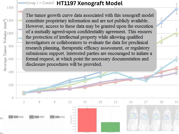
The HT1197 xenograft model reliably establishes tumors in immunodeficient mice, with a tumor take rate approaching 90% when 5 × 10^6 to 1 × 10^7 cells are injected subcutaneously in a matrix mixture such as Matrigel. Tumors typically become palpable within 10–14 days, with volumetric expansion occurring in a logarithmic pattern. Endpoint volumes of 1,200–1,500 mm³ are reached within 25–30 days post-inoculation under standard growth conditions.
While the model primarily supports subcutaneous implantation, orthotopic adaptation via intravesical delivery has been demonstrated in advanced research settings. In these configurations, the HT1197 model reproduces key features of urothelial wall invasion without extensive stromal penetration or distant metastasis, enabling the study of local progression and drug distribution within the bladder microenvironment.
Tumor morphology is generally cohesive, and vascular density supports adequate drug penetration and pharmacodynamic evaluation. The model is compatible with serial measurement protocols and is suitable for studies requiring tissue collection for histology, IHC, and biochemical assays. Bioluminescent or fluorescent labeling can be employed for longitudinal tracking when required.
Request a Custom Quote for HT1197 Xenograft ModelHistopathology and Immunohistochemical Profile
Histologically, HT1197 xenograft tumors form compact masses of polygonal epithelial cells with moderate nuclear pleomorphism, high nuclear-to-cytoplasmic ratios, and brisk mitotic activity. While not overtly differentiated, tumors preserve a degree of epithelial architecture, including cellular cohesion and intercellular junction formation. Necrotic zones are minimal, allowing for consistent tissue sampling.
Immunohistochemical staining reveals moderate to strong expression of E-cadherin, cytokeratin 7, and cytokeratin 18, confirming epithelial lineage. EGFR is intensely expressed at the membrane, and HER2/ERBB2 demonstrates patchy cytoplasmic and membranous staining. Ki-67 index is typically between 50–70%, indicating moderate to high proliferative capacity.
Vimentin expression is present in a subset of tumor cells, particularly near invasive fronts, suggesting partial EMT activation. MMP-9 is diffusely positive, while CD31 staining confirms moderate vascular density. Expression of uroplakin and CK20 is absent, corroborating the intermediate differentiation status of the model. Overall, the histological and immunophenotypic profile of HT1197 supports its classification as an epithelial-predominant, moderately aggressive bladder cancer model.
Preclinical Applications and Drug Response
The HT1197 xenograft model serves as a robust platform for evaluating therapeutic agents targeting EGFR, HER2, and downstream pathways such as PI3K/AKT and MAPK/ERK. Owing to its elevated EGFR expression, the model demonstrates partial responsiveness to tyrosine kinase inhibitors (TKIs) such as gefitinib and erlotinib, as well as to dual EGFR/HER2 inhibitors like lapatinib. Resistance tends to emerge through activation of alternative proliferative signaling pathways or incomplete suppression of receptor phosphorylation.
The model also exhibits sensitivity to DNA-damaging agents such as cisplatin and mitomycin C, albeit at doses higher than those effective in luminal-type models. This pharmacological profile mirrors clinical scenarios where receptor-overexpressing tumors show incomplete or transient chemotherapy responses. HDAC inhibitors and MMP-targeted therapies have also been tested successfully in this model, particularly in the context of preventing EMT progression and suppressing invasive phenotypes.
Given its intermediate epithelial-mesenchymal profile, HT1197 is frequently employed in mechanistic studies investigating partial EMT, reversible plasticity, and receptor-driven invasion. Its moderate growth kinetics and reliable take rate allow for well-controlled drug testing timelines and consistent endpoint analyses.
Request This Model
To request the HT1197 xenograft model or inquire about receptor-targeted therapy screening, EMT suppression studies, or customized orthotopic implementations, please contact us using the quote request form below. Our team is available to assist with study design, dosing schedules, and all aspects of model integration into your drug development program.
Request a Custom Quote for HT1197 Xenograft Model