SK-ES-1 Xenograft Model Overview
The SK-ES-1 xenograft model is derived from a human Ewing’s sarcoma cell line, SK-ES-1, which was established from a metastatic lesion in the femur of a patient diagnosed with Ewing’s sarcoma, a rare and highly aggressive cancer typically affecting children and adolescents. Ewing’s sarcoma is characterized by the presence of the EWS-FLI1 fusion gene, resulting from a chromosomal translocation between the EWSR1 and FLI1 genes. This fusion gene plays a crucial role in driving tumorigenesis, proliferation, and metastasis. The SK-ES-1 xenograft model is valuable for preclinical research, offering an ideal platform to study the biology of Ewing’s sarcoma, its metastatic behavior, and resistance to chemotherapy. Given the model’s ability to replicate key features of Ewing’s sarcoma, including tumor growth, metastasis, and EWS-FLI1 fusion protein expression, it is widely used to evaluate novel therapeutic strategies, including chemotherapy, targeted therapies, and immunotherapies.
Request a Custom Quote for SK-ES-1 Xenograft ModelBiological and Molecular Characteristics
SK-ES-1 cells are characterized by the presence of the EWS-FLI1 fusion gene, which is a hallmark of Ewing’s sarcoma and drives tumorigenesis. The fusion protein results from a translocation between the EWSR1 gene on chromosome 22 and the FLI1 gene on chromosome 11. This fusion protein acts as a transcription factor that alters the expression of genes involved in cell proliferation, survival, and differentiation, making EWS-FLI1 a key therapeutic target. SK-ES-1 cells also exhibit high expression of CD99, a membrane glycoprotein commonly expressed in Ewing’s sarcoma and used as a diagnostic marker. The model also shows activation of the PI3K/AKT and MAPK/ERK signaling pathways, which promote cell survival, proliferation, and metastasis, making the SK-ES-1 xenograft model useful for testing therapies targeting these pathways.
| Marker | Expression Level | Function |
|---|---|---|
| EWS-FLI1 Fusion | Present | Oncogenic fusion gene driving tumorigenesis |
| CD99 | High | Diagnostic marker for Ewing’s sarcoma |
| PI3K/AKT pathway | Dysregulated | Promotes cell survival and proliferation |
In Vivo Model Development and Tumorigenicity
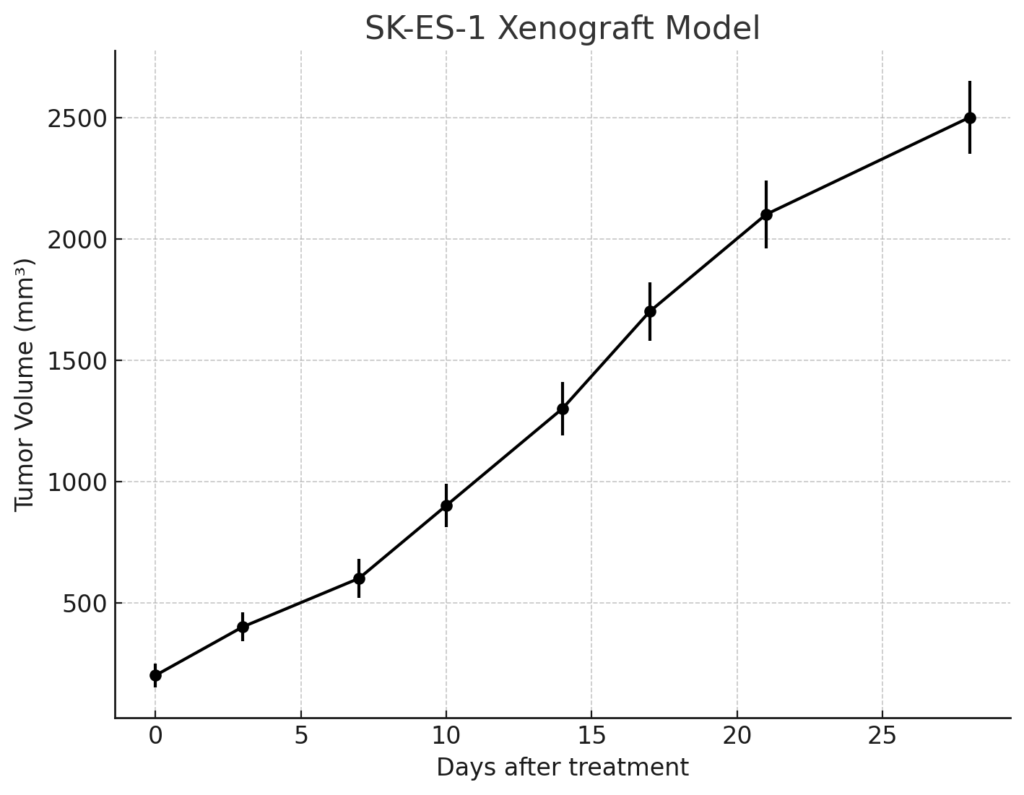
The SK-ES-1 xenograft model is typically established by subcutaneously implanting SK-ES-1 cells into immunocompromised mice, such as NOD/SCID or NSG mice, which lack functional T and B cells. Upon implantation, the cells form rapidly growing tumors that replicate key features of human Ewing’s sarcoma, including high cellularity, uniform morphology, and extensive vascularization. These tumors demonstrate significant mitotic activity and high levels of necrosis, reflecting the aggressive nature of the tumor. The SK-ES-1 model is commonly used to evaluate the effects of chemotherapy agents, such as vincristine, doxorubicin, and ifosfamide, which are part of the standard treatment regimen for Ewing’s sarcoma.
In addition to subcutaneous implantation, orthotopic models of SK-ES-1 can be established by implanting the cells into the bone or soft tissues of immunocompromised mice. This orthotopic model more accurately replicates the natural site of tumor growth and allows for the study of tumor progression, local invasion, and metastatic spread. The ability of SK-ES-1 cells to metastasize to distant organs, particularly the lungs and bones, makes this model highly relevant for studying metastatic disease and for evaluating therapies aimed at preventing or treating metastasis in Ewing’s sarcoma.
Request a Custom Quote for SK-ES-1 Xenograft ModelHistopathology and Immunohistochemical Profile
Histopathological examination of SK-ES-1 xenografts reveals the characteristic features of Ewing’s sarcoma, including small round blue cells with a high nuclear-to-cytoplasmic ratio, frequent mitotic figures, and areas of necrosis. Immunohistochemical staining of SK-ES-1 xenografts shows strong expression of CD99, a characteristic marker of Ewing’s sarcoma. The EWS-FLI1 fusion protein can be detected by specific probes, confirming its role in the tumor’s pathogenesis. The tumors also exhibit high levels of phosphorylated AKT, indicating the activation of the PI3K/AKT pathway, which supports tumor survival and growth. CD31 staining reveals significant angiogenesis, reflecting the tumor’s reliance on blood vessel formation for sustained growth.
Preclinical Applications and Drug Response
The SK-ES-1 xenograft model is widely used to evaluate the efficacy of chemotherapy agents such as vincristine, doxorubicin, and ifosfamide, which are the mainstays of treatment for Ewing’s sarcoma. Given its potential for developing resistance to chemotherapy, this model is highly valuable for studying chemotherapy resistance mechanisms and testing new agents aimed at overcoming these challenges. The SK-ES-1 model is also important for evaluating targeted therapies that inhibit the EWS-FLI1 fusion protein or modulate dysregulated signaling pathways such as PI3K/AKT and MAPK/ERK, which are involved in tumor survival and progression.
In addition to chemotherapy and targeted therapies, the SK-ES-1 xenograft model is increasingly used to evaluate the potential of immunotherapies, including immune checkpoint inhibitors and monoclonal antibodies targeting tumor-specific antigens. The model’s ability to replicate key features of Ewing’s sarcoma, including its aggressive growth, bone and soft tissue involvement, and metastatic potential, makes it an ideal platform for studying new treatment strategies. Furthermore, the ability of SK-ES-1 xenografts to metastasize to the lungs and bones provides an excellent opportunity to evaluate therapies aimed at preventing or treating metastatic disease in Ewing’s sarcoma.
Request This Model
To request the SK-ES-1 xenograft model for your preclinical studies, please use the form below. A customized quote and additional model specifications will be provided upon inquiry.
Request a Custom Quote for SK-ES-1 Xenograft Model