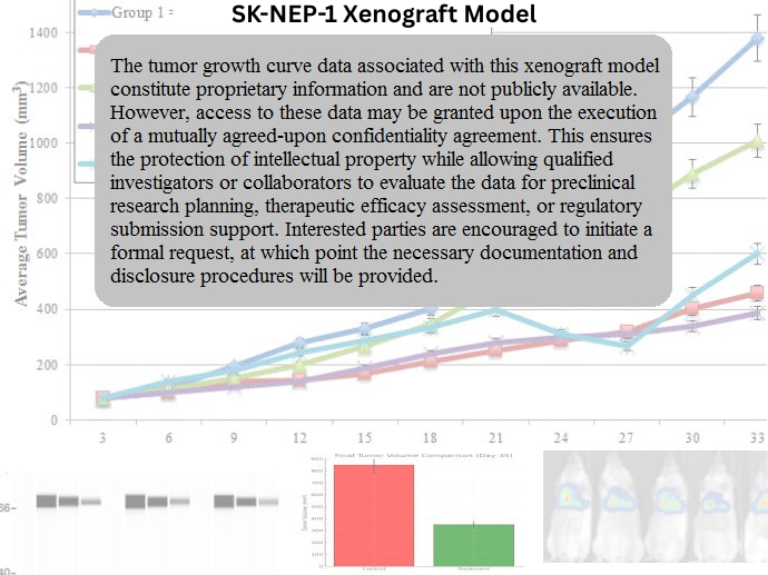
SK-NEP-1 Xenograft Model Overview
The SK-NEP-1 xenograft model is derived from a human renal cell carcinoma (RCC) cell line, SK-NEP-1, established from a metastatic renal tumor. RCC is one of the most common types of kidney cancer, and metastatic RCC remains a major cause of cancer-related mortality. The SK-NEP-1 model is particularly useful for studying clear cell renal carcinoma (ccRCC), which is the most common subtype of RCC, known for its high metastatic potential and resistance to traditional therapies. This xenograft model offers a valuable tool for evaluating the biology of renal cancer, tumor progression, metastasis, and testing therapeutic strategies aimed at improving the prognosis for RCC patients.
Request a Custom Quote for SK-NEP-1 Xenograft ModelBiological and Molecular Characteristics
SK-NEP-1 cells exhibit several features typical of renal cell carcinoma, including epithelial cell markers such as cytokeratins and epithelial membrane antigen (EMA). These cells are particularly valuable for studying ccRCC as they harbor mutations in the von Hippel-Lindau (VHL) gene, a common mutation found in ccRCC that leads to the accumulation of hypoxia-inducible factors (HIFs). The upregulation of HIFs results in the activation of downstream target genes such as vascular endothelial growth factor (VEGF), which plays a key role in tumor angiogenesis and progression. The model also shows alterations in the PI3K/AKT and MAPK signaling pathways, contributing to tumor cell survival, proliferation, and metastasis. These molecular features make the SK-NEP-1 xenograft model particularly useful for studying therapeutic strategies targeting angiogenesis and aberrant signaling in RCC.
| Marker | Expression Level | Function |
|---|---|---|
| Cytokeratin | High | Epithelial cell marker |
| EMA | High | Epithelial membrane antigen |
| VHL | Mutated | Tumor suppressor gene regulating hypoxia response |
| HIF-1α/HIF-2α | Elevated | Transcription factors promoting angiogenesis |
| VEGF | Elevated | Angiogenesis factor promoting tumor growth |
In Vivo Model Development and Tumorigenicity
The SK-NEP-1 xenograft model is typically established by subcutaneously implanting SK-NEP-1 cells into immunocompromised mice, such as NOD/SCID or NSG mice, which lack functional T and B cells. Upon implantation, the cells form solid tumors that closely replicate the clinical features of human renal cell carcinoma, including rapid growth, necrosis, and significant vascularization. The model is commonly used to evaluate the effects of chemotherapy and targeted therapies, such as VEGF inhibitors, which target the tumor’s angiogenic properties. Given its ability to develop resistance to conventional therapies, the SK-NEP-1 model is particularly valuable for studying resistance mechanisms and for evaluating combination therapies that aim to enhance treatment efficacy.
In addition to subcutaneous implantation, orthotopic models of SK-NEP-1 can be established by implanting the cells directly into the kidneys of immunocompromised mice. This orthotopic model more accurately replicates the natural site of tumor growth and allows for the study of tumor progression, local invasion, and metastasis, which is a key feature of RCC. The ability of SK-NEP-1 tumors to metastasize to distant organs, such as the lungs, liver, and bones, makes this model ideal for testing therapies aimed at preventing or treating metastasis in RCC.
Request a Custom Quote for SK-NEP-1 Xenograft ModelHistopathology and Immunohistochemical Profile
Histopathological examination of SK-NEP-1 xenografts reveals characteristic features of renal cell carcinoma, including irregular glandular structures, areas of necrosis, and a high mitotic index. Immunohistochemical staining of SK-NEP-1 xenografts shows strong expression of epithelial markers such as cytokeratin and EMA, confirming the epithelial origin of the tumor. Additionally, high levels of VEGF and HIF-1α/HIF-2α are observed, reflecting the dysregulation of the VHL gene and the upregulation of key angiogenic factors. The tumors also exhibit high levels of phosphorylated AKT, indicating activation of the PI3K/AKT pathway, which promotes cell survival and resistance to apoptosis. CD31 staining reveals significant vascularization, highlighting the tumor’s reliance on angiogenesis for sustained growth.
Preclinical Applications and Drug Response
The SK-NEP-1 xenograft model is widely used to evaluate the efficacy of various therapeutic agents, particularly those targeting the angiogenesis pathway, such as VEGF inhibitors like bevacizumab. The model is also valuable for studying the effects of standard RCC therapies, such as tyrosine kinase inhibitors (e.g., sunitinib and sorafenib), which target key growth factor receptors involved in tumor progression and angiogenesis. The SK-NEP-1 xenograft model is also employed to evaluate novel therapies that aim to restore VHL function or inhibit HIF-1α and HIF-2α, as these pathways are central to the pathogenesis of RCC.
In addition to targeted therapies, the SK-NEP-1 xenograft model is useful for investigating combination therapies that include chemotherapy agents and novel targeted treatments. The model’s ability to replicate the features of RCC, including its aggressive nature, angiogenesis, and metastatic potential, makes it an ideal platform for testing new therapeutic strategies in preclinical studies. The model is also valuable for studying the tumor microenvironment, including immune cell interactions, which are essential for understanding resistance to treatment and improving the effectiveness of immunotherapies.
Request This Model
To request the SK-NEP-1 xenograft model for your preclinical studies, please use the form below. A customized quote and additional model specifications will be provided upon inquiry.
Request a Custom Quote for SK-NEP-1 Xenograft Model