Hs 766T Xenograft Model Overview
The Hs 766T xenograft model is derived from a human pancreatic cancer cell line, Hs 766T, established from a patient with pancreatic ductal adenocarcinoma (PDAC). PDAC is a highly aggressive and lethal cancer with a poor prognosis, primarily due to its late-stage diagnosis, rapid metastasis, and resistance to most therapies. The Hs 766T xenograft model is invaluable for preclinical research, providing insights into pancreatic cancer biology, tumor progression, and metastasis. This model is widely used for evaluating the efficacy of chemotherapy, targeted therapies, and immunotherapies aimed at overcoming the intrinsic resistance of pancreatic tumors.
Request a Custom Quote for Hs 766T Xenograft ModelBiological and Molecular Characteristics
Hs 766T cells are characterized by their epithelial origin, expressing common markers associated with pancreatic cancer, such as cytokeratins, epithelial membrane antigen (EMA), and the pancreatic cancer-associated antigen CA19-9. These cells harbor mutations in the KRAS oncogene, which is found in over 90% of PDAC cases and contributes to tumorigenesis, survival, and metastasis. Additionally, Hs 766T cells show dysregulated signaling in pathways such as PI3K/AKT and MAPK/ERK, which promote cell survival, proliferation, and metastasis. The model also exhibits loss of the tumor suppressor gene TP53, which contributes to the uncontrolled proliferation and resistance to apoptosis commonly seen in pancreatic cancer. Given these molecular characteristics, the Hs 766T xenograft model is useful for testing therapies targeting KRAS, PI3K/AKT, and MAPK pathways, as well as therapies aimed at restoring p53 function.
| Marker | Expression Level | Function |
|---|---|---|
| Cytokeratin | High | Epithelial cell marker |
| EMA | High | Epithelial membrane antigen |
| CA19-9 | Elevated | Pancreatic cancer biomarker |
| KRAS | Mutated | Oncogene involved in tumor progression |
| PI3K/AKT pathway | Dysregulated | Promotes cell survival and proliferation |
In Vivo Model Development and Tumorigenicity
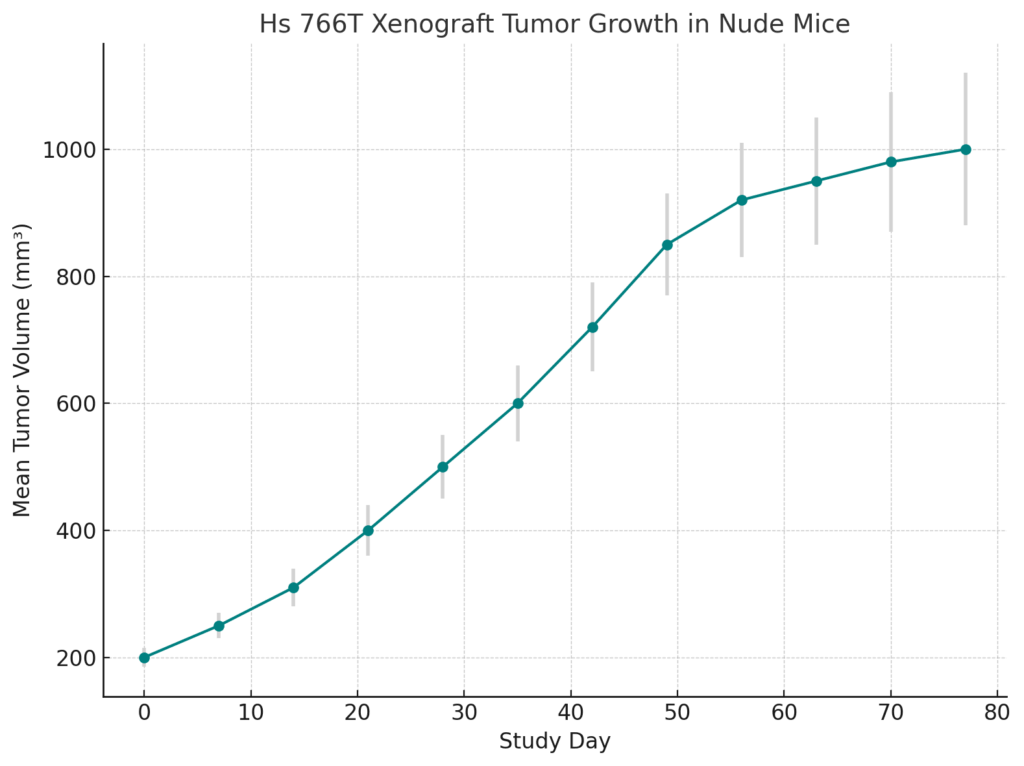
The Hs 766T xenograft model is typically established by subcutaneously implanting Hs 766T cells into immunocompromised mice, such as NOD/SCID or NSG mice, which lack functional T and B cells. Once implanted, the cells form tumors that closely resemble human pancreatic ductal adenocarcinoma in terms of histology, growth, and metastasis. These tumors exhibit high cellularity, necrosis, and significant vascularization, which are characteristic of rapidly growing pancreatic cancer. The Hs 766T model is particularly valuable for evaluating the efficacy of chemotherapy agents, such as gemcitabine and cisplatin, as well as for investigating the molecular mechanisms underlying chemotherapy resistance.
In addition to subcutaneous implantation, orthotopic implantation of Hs 766T cells into the pancreas of immunocompromised mice can be performed. This orthotopic model closely mimics the natural progression of pancreatic cancer, including local invasion and metastatic spread to distant organs, such as the liver, lungs, and peritoneum. This more clinically relevant model allows for the study of pancreatic cancer progression and provides an excellent platform for evaluating treatments that target both primary tumors and metastases.
Request a Custom Quote for Hs 766T Xenograft ModelHistopathology and Immunohistochemical Profile
Histopathological examination of Hs 766T xenografts reveals the characteristic features of pancreatic ductal adenocarcinoma, including irregular glandular structures, high cellularity, and areas of necrosis. The tumors exhibit pleomorphic cells with abundant cytoplasm, high mitotic activity, and irregular nuclear morphology, indicative of aggressive tumor growth. Immunohistochemical staining of Hs 766T xenografts shows strong expression of epithelial markers, such as cytokeratins and EMA, confirming the epithelial origin of the tumor. Elevated levels of CA19-9 are also detected, which is consistent with the biomarker’s role in pancreatic cancer. Additionally, the tumors show high levels of phosphorylated AKT, indicating activation of the PI3K/AKT pathway, a key signaling pathway involved in tumor survival and proliferation. The tumors are also highly vascularized, as assessed by CD31 staining, reflecting the angiogenic potential of the tumors, which supports their rapid growth.
Preclinical Applications and Drug Response
The Hs 766T xenograft model is widely used to evaluate the efficacy of chemotherapy agents, particularly gemcitabine, cisplatin, and other platinum-based drugs. Given its sensitivity to these agents, the model is valuable for testing new chemotherapy agents or combination therapies aimed at overcoming chemotherapy resistance. The model is also useful for evaluating targeted therapies, particularly those that inhibit the KRAS, PI3K/AKT, and MAPK/ERK signaling pathways, which are commonly dysregulated in pancreatic cancer.
In addition to chemotherapy and targeted therapies, the Hs 766T xenograft model is frequently used to evaluate the effectiveness of immunotherapies, including immune checkpoint inhibitors and monoclonal antibodies targeting cancer-specific antigens, such as CA19-9. The model’s ability to replicate key features of pancreatic cancer, including its high metastatic potential and resistance to therapy, makes it an ideal platform for studying new therapeutic agents. Furthermore, Hs 766T xenografts are increasingly used to investigate combination therapies that include chemotherapy and novel immunotherapies or targeted inhibitors to improve treatment efficacy and overcome drug resistance.
Request This Model
To request the Hs 766T xenograft model for your preclinical studies, please use the form below. A customized quote and additional model specifications will be provided upon inquiry.
Request a Custom Quote for Hs 766T Xenograft Model