HEL 92.1.7 Xenograft Model Overview
The HEL 92.1.7 xenograft model is derived from a human erythroleukemia cell line originally isolated from the peripheral blood of a 30-year-old male with acute erythroid leukemia (M6 subtype under the FAB classification). This model exhibits characteristics of erythroid/myeloid lineage malignancies and retains many features of early-stage erythroid precursor cells. HEL 92.1.7 xenografts offer a valuable in vivo platform for studying erythroleukemia pathophysiology, erythropoiesis, and JAK-STAT pathway dysregulation, particularly in the context of myeloproliferative neoplasms (MPNs) and acute erythroid leukemia. Due to their constitutive JAK2 V617F mutation and robust engraftment potential in immunocompromised mice, these xenografts are widely utilized for evaluating kinase inhibitors, epigenetic therapies, and cytotoxic regimens targeting erythroid leukemia.
Request a Custom Quote for HEL 92.1.7 Xenograft ModelBiological and Molecular Characteristics
HEL 92.1.7 cells exhibit a loosely adherent, round-to-oval morphology and express markers indicative of erythroid and myeloid lineage differentiation, including glycophorin A, CD71 (transferrin receptor), and low levels of CD13 and CD33. A defining feature of this model is the presence of the JAK2 V617F mutation, which leads to constitutive activation of the JAK-STAT signaling pathway, promoting abnormal erythroid proliferation and survival. The cell line expresses high levels of phosphorylated STAT5 and downstream effectors, along with disrupted cell cycle control involving p21 and p27. The HEL 92.1.7 genome is complex and exhibits chromosomal abnormalities including trisomy 8 and other structural rearrangements consistent with leukemic karyotypes. The model is p53 wild-type and BCR-ABL negative.
| Characteristic | HEL 92.1.7 Cell Line Profile |
|---|---|
| Disease Origin | Acute erythroid leukemia (FAB M6) |
| JAK2 Status | V617F mutated |
| Lineage Markers | CD71, glycophorin A, CD13, CD33 |
| BCR-ABL Status | Negative |
| p53 Status | Wild-type |
| Signaling Pathways | Activated JAK-STAT |
In Vivo Model Development and Tumorigenicity
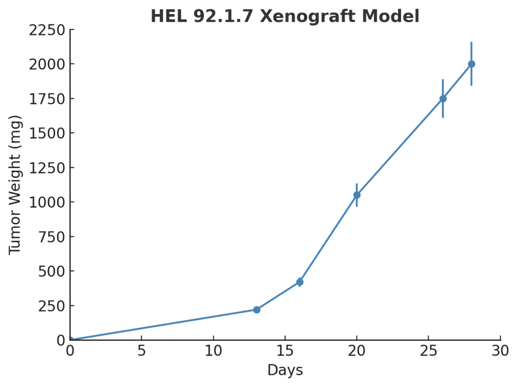
The HEL 92.1.7 xenograft model is developed by intravenous or subcutaneous injection of cultured cells into immunodeficient murine hosts, typically NOD/SCID or NSG strains. In subcutaneous settings, tumor formation occurs within 10–14 days post-injection and progresses to 700–900 mm³ over 4–5 weeks, exhibiting dense, infiltrative growth. In disseminated models, leukemic infiltration is observed in the bone marrow, spleen, and liver, mimicking systemic erythroleukemia. Tumor take rates are consistently high, and the JAK2-mutant background ensures pathway-specific reproducibility for drug screening. These models are especially well suited for evaluating the in vivo effects of JAK inhibitors, epigenetic modifiers, and agents modulating erythroid differentiation or proliferation.
Request a Custom Quote for HEL 92.1.7 Xenograft ModelHistopathology and Immunohistochemical Profile
Histological examination of HEL 92.1.7 xenografts shows sheets of monomorphic round cells with basophilic cytoplasm, high nuclear-to-cytoplasmic ratio, and frequent mitotic figures. H&E staining highlights erythroid-like morphology with scattered apoptotic bodies and areas of necrosis in larger tumors. In systemic models, infiltration of neoplastic cells into hematopoietic organs replicates clinical features of acute erythroid leukemia. Immunohistochemically, xenograft tissues show strong expression of CD71 and glycophorin A, confirming erythroid lineage, along with phosphorylated STAT5, indicating active JAK-STAT signaling. CD45 expression is typically low, differentiating the model from lymphoid leukemias.
Preclinical Applications and Drug Response
The HEL 92.1.7 xenograft model is widely employed in preclinical testing of JAK2 inhibitors (e.g., ruxolitinib, fedratinib), particularly in the context of JAK2-mutated myeloproliferative disorders and erythroleukemia. It serves as a valuable tool for evaluating agents that induce erythroid differentiation or apoptosis, as well as small molecules targeting downstream STAT5 signaling, cell cycle checkpoints, and epigenetic regulators such as HDACs and DNMTs. The model also supports studies of combination therapies involving kinase inhibition and DNA-damaging agents. Its reproducible tumor growth, defined molecular profile, and systemic infiltration capabilities make HEL 92.1.7 an essential model for translational leukemia research and hematologic drug development.
Request This Model
To utilize the HEL 92.1.7 xenograft model for erythroleukemia research or evaluation of JAK2-targeted therapies, contact our scientific team to obtain detailed model parameters and support for custom in vivo study design tailored to hematologic malignancy development and drug response profiling.
Request a Custom Quote for HEL 92.1.7 Xenograft Model