LS180 Xenograft Model Overview
The LS180 xenograft model is derived from a human colorectal adenocarcinoma cell line originally established from a metastatic lesion in the left supraclavicular region of a 58-year-old female. As a representative model of metastatic colorectal cancer (CRC), LS180 is frequently utilized in preclinical oncology studies due to its epithelial differentiation, stable growth kinetics in immunodeficient mice, and clinically relevant mutation profile. The cell line displays genetic characteristics consistent with chromosomal instability, one of the predominant molecular subtypes of CRC. This model is especially valuable in testing the efficacy of targeted therapeutics and pathway inhibitors in KRAS-mutant CRC, and it supports translational investigations that require a reproducible, genetically defined, and histopathologically faithful in vivo system.
Request a Custom Quote for LS180 Xenograft ModelBiological and Molecular Characteristics
LS180 cells are moderately to well-differentiated and maintain epithelial polarity, expressing tight junction proteins and cell adhesion markers such as E-cadherin. The cell line is characterized by a KRAS G13D mutation, a clinically relevant alteration that imparts resistance to EGFR-targeted therapies while maintaining responsiveness to downstream inhibition of the MAPK and PI3K pathways. Unlike many colorectal cancer models, LS180 cells retain wild-type TP53 and are microsatellite stable (MSS), making them suitable for evaluating non-MSI-driven therapeutic interventions. The cells also express high levels of carcinoembryonic antigen (CEA), a clinically significant tumor marker in CRC. These molecular attributes enable LS180 to serve as a precise model for genotype-stratified drug testing and mechanism-of-action studies in colorectal oncology.
| Characteristic | LS180 Cell Line Profile |
|---|---|
| Tissue of Origin | Colorectal adenocarcinoma (metastatic) |
| KRAS Status | Mutant (G13D) |
| TP53 Status | Wild-type |
| CEA Expression | High |
| Growth Characteristics | Adherent, epithelial morphology |
| MSI Status | Microsatellite stable (MSS) |
In Vivo Model Development and Tumorigenicity
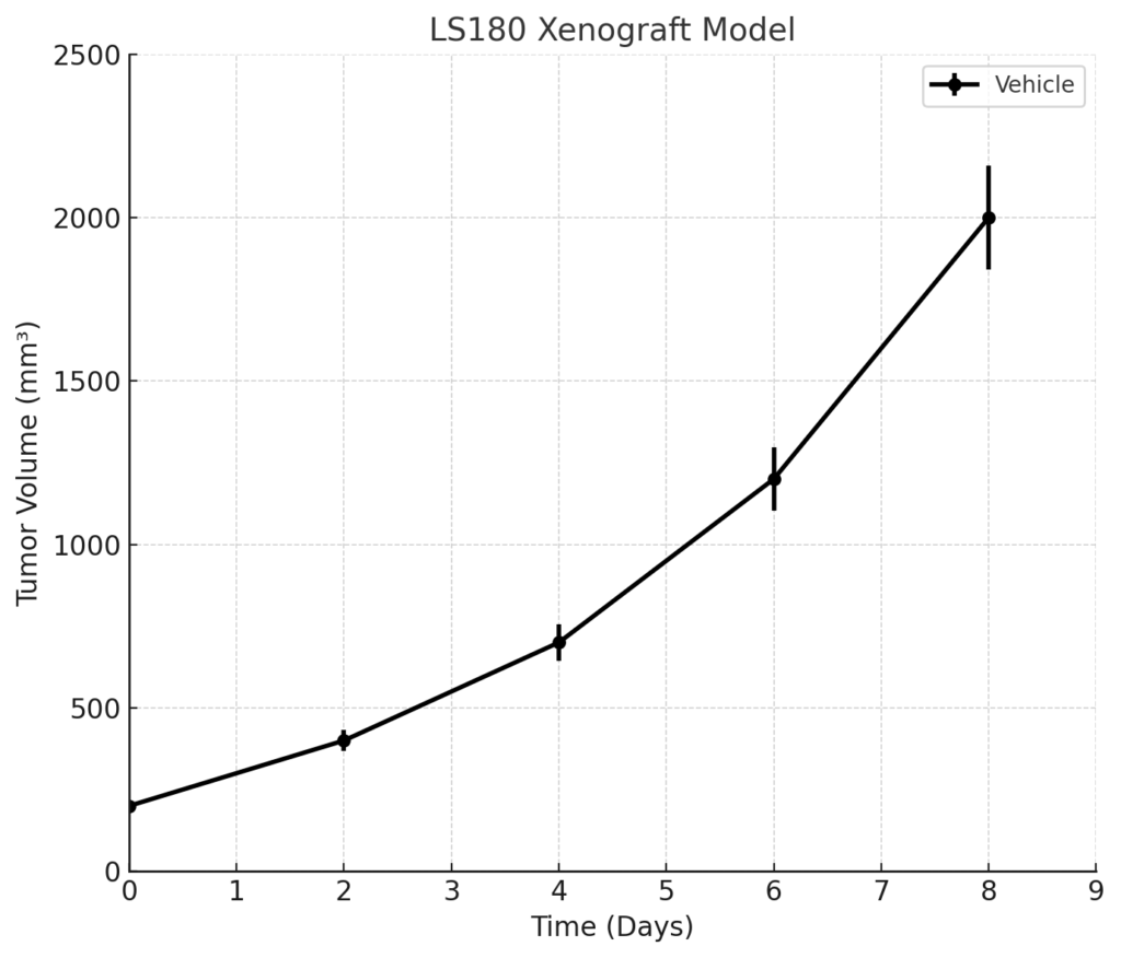
Xenograft formation with LS180 cells is typically achieved through subcutaneous injection into immunodeficient murine hosts, including athymic nude or NOD/SCID mice. Tumor engraftment is robust, with initial tumor nodules detectable within 7 to 10 days post-inoculation. LS180 xenografts exhibit consistent exponential growth, often reaching target volumes between 700 and 900 mm³ within 4 to 5 weeks. The model offers high reproducibility across cohorts, allowing for efficient comparison of treatment arms in multi-arm preclinical efficacy studies. Because the KRAS G13D mutation drives tumorigenesis in this model, it is particularly useful in evaluating drugs that bypass upstream receptor blockade in favor of direct inhibition of RAS signaling effectors.
Request a Custom Quote for LS180 Xenograft ModelHistopathology and Immunohistochemical Profile
LS180 xenografts present as moderately differentiated adenocarcinomas with glandular morphology, luminal necrosis, and mucin production consistent with primary colorectal tumors. Histologically, the tumors retain features of epithelial polarity and cohesion, mirroring the differentiation status of the originating human cancer. Immunohistochemical staining reveals strong expression of CEA, cytokeratin 20 (CK20), and epithelial membrane antigen (EMA), confirming intestinal lineage. E-cadherin and β-catenin are preserved, supporting intact adherens junction architecture. Vascularization within the tumor microenvironment is moderate, and stromal infiltration remains limited, which contributes to the model’s reproducibility and phenotypic stability under therapeutic pressure.
Preclinical Applications and Drug Response
The LS180 xenograft model is widely applied in the evaluation of therapeutic agents for KRAS-mutant CRC. Due to its G13D mutation, it demonstrates resistance to anti-EGFR monoclonal antibodies such as cetuximab, providing a clinically relevant platform for studying alternative therapeutic strategies. MEK, PI3K, and mTOR inhibitors have demonstrated measurable antitumor activity in LS180 xenografts, particularly in rational combination regimens. This model is also employed in studies of drug penetration and retention, biomarker expression changes, and tumor growth delay following treatment with small molecules, RNA-based therapeutics, and antibody-drug conjugates. The preserved wild-type TP53 status allows for selective testing of agents targeting intact apoptotic signaling, offering an additional layer of translational insight.
Request This Model
To incorporate the LS180 xenograft model into your preclinical research pipeline, initiate a consultation to discuss custom study design, model availability, and service specifications.
Request a Custom Quote for LS180 Xenograft Model