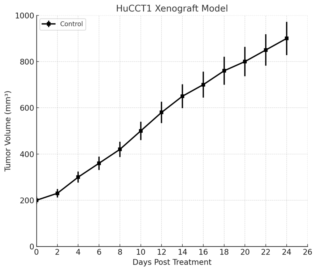
HuCCT1 Xenograft Model Overview
The HuCCT1 xenograft model is derived from a human intrahepatic cholangiocarcinoma (iCCA), a malignancy of the bile duct epithelium within the liver. Originally established from the bile duct tissue of a 56-year-old Japanese patient, the HuCCT1 cell line is widely recognized as a representative model of primary liver tumors with glandular differentiation and aggressive growth behavior. Given the rarity and poor prognosis of iCCA, the HuCCT1 xenograft model serves as a valuable platform for studying tumor biology, evaluating therapeutic responses, and dissecting the molecular signaling pathways implicated in biliary carcinogenesis.
This model is particularly suitable for preclinical investigation of agents targeting FGFR alterations, KRAS mutations, and the tumor stroma, which is prominent in cholangiocarcinoma pathogenesis. It supports both subcutaneous and orthotopic tumor implantation and is compatible with imaging modalities, making it ideal for translational studies involving targeted therapies and immunomodulation.
Request a Custom Quote for HuCCT1 Xenograft ModelBiological and Molecular Characteristics
HuCCT1 cells exhibit an epithelial phenotype with strong cell-cell adhesion, glandular differentiation, and formation of tubular structures in vitro. The model harbors mutations in KRAS (G12D), which drive constitutive activation of the MAPK/ERK pathway. Unlike many cholangiocarcinoma models, HuCCT1 is wild-type for FGFR2 fusions but still exhibits sensitivity to FGFR inhibitors through pathway crosstalk. It also expresses key epithelial and biliary markers, such as CK7, CK19, and EpCAM.
| Characteristic | HuCCT1 Profile |
|---|---|
| Tumor Type | Intrahepatic cholangiocarcinoma (iCCA) |
| KRAS Status | Mutant (G12D) |
| FGFR2 Fusion | Negative |
| IDH1/2 Mutation | Wild-type |
| CK7 / CK19 Expression | High |
| EpCAM | Positive |
| E-cadherin | Retained |
| TP53 | Wild-type |
| Tumor Growth Rate | Moderate to high |
| Invasiveness | High (locally and systemically in orthotopic models) |
These molecular characteristics make HuCCT1 suitable for modeling KRAS-driven biliary cancers and for screening targeted therapies with relevance to iCCA.
In Vivo Model Development and Tumorigenicity
HuCCT1 xenografts are most commonly established via subcutaneous injection of 5 × 10⁶ to 1 × 10⁷ cells into the flank of immunodeficient mice such as NOD/SCID or nude mice. Tumor take rates typically exceed 90%, and growth is relatively consistent, with tumors reaching 1,000 mm³ in 4–6 weeks depending on host strain and site of injection.
For more physiologically relevant modeling, orthotopic implantation into the liver can be performed by injecting a suspension of cells into the left hepatic lobe under surgical guidance. This approach more accurately recapitulates the tumor microenvironment, including liver-specific vasculature, bile duct architecture, and stromal interaction. Orthotopic tumors can invade into portal areas and form intrahepatic metastases, mimicking advanced iCCA.
The HuCCT1 model is compatible with bioluminescent imaging (luciferase-tagged lines) and allows real-time monitoring of tumor growth, drug response, and metastatic dissemination.
Request a Custom Quote for HuCCT1 Xenograft ModelHistopathology and Immunohistochemical Profile
HuCCT1 xenografts display histologic features of well to moderately differentiated adenocarcinoma. Tumors grow in glandular or duct-like patterns with mucinous cytoplasm and desmoplastic stroma. Tumor cells exhibit moderate pleomorphism, mitotic activity, and variable nuclear polarity.
Immunohistochemically, tumors are positive for cytokeratin 7 (CK7), cytokeratin 19 (CK19), EpCAM, and E-cadherin. Ki-67 proliferation indices range from 40% to 60%, depending on tumor region and host immune context. Phospho-ERK1/2 staining is consistently elevated due to KRAS activation. Vascular and stromal markers such as CD31 and α-SMA highlight the desmoplastic and angiogenic components of the tumor microenvironment.
These features support the utility of HuCCT1 xenografts for biomarker-driven therapeutic studies and for evaluating drug penetration and stromal remodeling in fibrotic tumor environments.
Preclinical Applications and Drug Response
The HuCCT1 xenograft model is extensively employed in cholangiocarcinoma research due to its molecular fidelity and predictable in vivo behavior. It is particularly valuable for:
- KRAS pathway inhibition using MEK or ERK inhibitors
- Anti-stromal therapies, including TGF-β pathway inhibitors and stroma-targeted nanoparticles
- FGFR and VEGF pathway blockade, even in the absence of FGFR fusions, due to crosstalk and paracrine signaling
- DNA-damaging agents, such as gemcitabine, cisplatin, and oxaliplatin
- Immune checkpoint blockade in humanized or partially immunocompetent mouse strains
- Combination regimens, such as MEK inhibitors with anti-fibrotic agents or angiogenesis inhibitors
This model also supports pharmacodynamic studies involving ERK phosphorylation, tumor fibrosis reduction, and apoptosis quantification, making it suitable for both early-stage and late-stage drug evaluation in biliary tract cancers.
Request This Model
To incorporate the HuCCT1 xenograft model into your intrahepatic cholangiocarcinoma study or targeted therapy development pipeline, request a tailored xenograft services package below. Offerings include orthotopic implantation, drug response profiling, stroma modulation studies, and advanced histopathology.
Request a Custom Quote for HuCCT1 Xenograft Model