Daoy Xenograft Model Overview
The Daoy xenograft model is derived from a human medulloblastoma and is widely used in preclinical neuro-oncology research due to its reproducible tumor formation, moderate growth kinetics, and responsiveness to central nervous system (CNS)-targeted therapies. Originally isolated from the cerebellum of a 4-year-old male patient, the Daoy cell line represents a SHH (Sonic Hedgehog) subtype of medulloblastoma, which is one of the four major molecular classifications of this pediatric brain tumor. Daoy xenografts are particularly valuable for studies focused on SHH pathway modulation, cerebellar tumorigenesis, and radiation sensitization strategies.
Although Daoy is less aggressive than MYC-amplified Group 3 models such as D283, it retains essential characteristics of high-risk medulloblastoma, including sustained proliferation, p53 dysfunction, and anchorage-independent growth. Its compatibility with orthotopic implantation and relatively stable phenotype makes it an ideal system for mechanistic studies of cerebellar tumor biology, genetic perturbation screening, and CNS drug penetration.
Request a Custom Quote for Daoy Xenograft ModelBiological and Molecular Characteristics
The Daoy cell line demonstrates molecular features typical of SHH-driven medulloblastoma, including high expression of GLI1, PTCH1, and SFRP1, all of which are downstream effectors of the SHH signaling pathway. Importantly, the model lacks MYC or MYCN amplification, differentiating it from more aggressive medulloblastoma variants. The TP53 gene in Daoy is mutated, which impairs canonical apoptosis and cell cycle control, contributing to modest resistance to genotoxic stress.
Daoy expresses neural progenitor markers such as nestin, SOX2, and β-III tubulin, confirming its neuroectodermal origin. The cell line also displays moderate levels of BCL-2, cyclin D2, and CDK6, indicative of proliferative capacity without extensive dedifferentiation. Active AKT and ERK signaling further supports tumor cell survival under stress conditions. Chromosomal analysis shows relative stability, and the line is karyotypically near-diploid.
| Characteristic | Daoy Profile |
|---|---|
| Tumor Type | Human medulloblastoma (SHH subtype) |
| Origin | Cerebellum, 4-year-old male patient |
| TP53 Status | Mutant |
| MYC/MYCN Amplification | Absent |
| SHH Pathway Markers | GLI1+, PTCH1+, SFRP1+ |
| Neural Markers | Nestin+, β-III Tubulin+, SOX2+ |
| Cell Cycle Regulators | Cyclin D2+, CDK6+ |
| Apoptosis Modulators | BCL-2+, Survivin+ |
| PI3K/AKT and MAPK Activity | Elevated |
| Morphology | Neuroepithelial, polygonal |
This profile supports the model’s role in studying SHH signaling, tumor cell differentiation, and radiation response mechanisms in cerebellar tumors.
In Vivo Model Development and Tumorigenicity
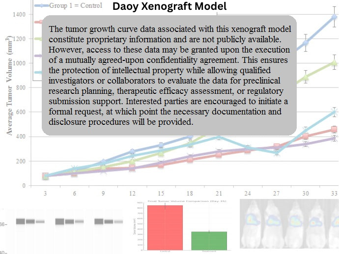
Daoy xenografts are commonly established via subcutaneous or orthotopic implantation in immunodeficient mice. Subcutaneous inoculation of 5 × 10^6 to 1 × 10^7 cells in Matrigel yields tumors with take rates exceeding 90%, becoming palpable within 10–12 days and reaching volumes of 1,000–1,300 mm³ by week 4. Tumor growth is consistent and moderately paced, making the model suitable for long-term treatment studies and schedule-dependent drug evaluation.
Orthotopic implantation into the cerebellum offers anatomical accuracy and has been employed to replicate localized brain tumor formation, vascular engagement, and SHH microenvironmental dependencies. While orthotopic Daoy models do not exhibit spontaneous metastasis, they enable high-resolution evaluation of drug bioavailability across the blood–brain barrier (BBB), as well as radiation and surgical intervention strategies.
The Daoy model is also suitable for luciferase or GFP tagging to facilitate non-invasive bioluminescence imaging, cell lineage tracing, and intravital microscopy in orthotopic settings.
Request a Custom Quote for Daoy Xenograft ModelHistopathology and Immunohistochemical Profile
Histological analysis of Daoy xenograft tumors reveals solid sheets of neuroepithelial cells with moderate nuclear pleomorphism, high nuclear-to-cytoplasmic ratio, and occasional formation of Homer Wright rosettes. Tumors are cellular with low stromal content and minimal necrosis under standard growth conditions.
Immunohistochemistry demonstrates strong expression of Ki-67 (40–60%), β-III tubulin, and SOX2, confirming proliferative potential and neural progenitor identity. Nuclear positivity for GLI1 and PTCH1 supports active SHH signaling, while BCL-2 and survivin contribute to apoptotic resistance. CD31 staining reveals moderate vascularization, with well-formed microvasculature distributed evenly throughout the tumor.
Daoy xenografts are negative for MYC, GFAP, and markers of glial differentiation, reinforcing their neuroepithelial lineage. Phospho-AKT and phospho-ERK are localized to cytoplasmic compartments, consistent with activated survival pathways.
Preclinical Applications and Drug Response
The Daoy xenograft model is extensively used for testing SHH pathway inhibitors, including SMO antagonists (e.g., vismodegib, sonidegib) and GLI transcriptional blockers. It is also an important tool for studying radiation sensitization, as its TP53 mutation results in impaired DNA damage response but retains partial susceptibility to apoptosis under combination therapy conditions.
Daoy tumors are moderately sensitive to cisplatin, etoposide, and irinotecan, although resistance develops over time. The model is well suited for screening epigenetic agents such as HDAC inhibitors, EZH2 inhibitors, and BET bromodomain inhibitors, which have shown differentiation-inducing effects. Its use in orthotopic models supports assessment of drug pharmacokinetics and penetration into the cerebellar parenchyma.
Due to its relatively moderate growth kinetics and anatomical relevance, Daoy is frequently employed in studies of maintenance therapy, residual disease modeling, and radiotherapy optimization. It is also used for CRISPR/Cas9-based functional screening of SHH-regulated genes and tumor suppressors.
Request This Model
To request the Daoy xenograft model or integrate it into studies of medulloblastoma biology, SHH pathway inhibition, or CNS-targeted therapy evaluation, please use the quote request form below. We provide support for subcutaneous and orthotopic implantation protocols, imaging setup, and longitudinal therapeutic studies.
Request a Custom Quote for Daoy Xenograft Model